ARG55041
anti-Ki-67 antibody [Ki-67] (PE)
anti-Ki-67 antibody [Ki-67] (PE) for Flow cytometry and Human,Cow
Overview
| Product Description | PE-conjugated Mouse Monoclonal antibody [Ki-67] recognizes Ki-67 |
|---|---|
| Tested Reactivity | Hu, Cow |
| Tested Application | FACS |
| Specificity | The mouse monoclonal antibody Ki-67 recognizes Ki-67 antigen, a non-histone nuclear protein expressed exclusively in proliferating cells. |
| Host | Mouse |
| Clonality | Monoclonal |
| Clone | Ki-67 |
| Isotype | IgG1 |
| Target Name | Ki-67 |
| Antigen Species | Human |
| Immunogen | Nuclei of the Hodgkin lymphoma cell line L428. |
| Conjugation | PE |
| Alternate Names | Antigen KI-67; MIB-; KIA; MIB-1; PPP1R105 |
Application Instructions
| Application Suggestion |
|
||||
|---|---|---|---|---|---|
| Application Note | * The dilutions indicate recommended starting dilutions and the optimal dilutions or concentrations should be determined by the scientist. |
Properties
| Form | Liquid |
|---|---|
| Purification | Purified |
| Buffer | PBS and 15 mM Sodium azide. |
| Preservative | 15 mM Sodium azide |
| Storage Instruction | Aliquot and store in the dark at 2-8°C. Keep protected from prolonged exposure to light. Avoid repeated freeze/thaw cycles. Suggest spin the vial prior to opening. The antibody solution should be gently mixed before use. |
| Note | For laboratory research only, not for drug, diagnostic or other use. |
Bioinformation
| Database Links | |
|---|---|
| Gene Symbol | MKI67 |
| Gene Full Name | marker of proliferation Ki-67 |
| Background | This gene encodes a nuclear protein that is associated with and may be necessary for cellular proliferation. Alternatively spliced transcript variants have been described. A related pseudogene exists on chromosome X. [provided by RefSeq, Mar 2009] |
| Function | Required to maintain individual mitotic chromosomes dispersed in the cytoplasm following nuclear envelope disassembly (PubMed:27362226). Associates with the surface of the mitotic chromosome, the perichromosomal layer, and covers a substantial fraction of the chromosome surface (PubMed:27362226). Prevents chromosomes from collapsing into a single chromatin mass by forming a steric and electrostatic charge barrier: the protein has a high net electrical charge and acts as a surfactant, dispersing chromosomes and enabling independent chromosome motility (PubMed:27362226). Binds DNA, with a preference for supercoiled DNA and AT-rich DNA (PubMed:10878551). Does not contribute to the internal structure of mitotic chromosomes (By similarity). May play a role in chromatin organization (PubMed:24867636). It is however unclear whether it plays a direct role in chromatin organization or whether it is an indirect consequence of its function in maintaining mitotic chromosomes dispersed (Probable). [UniProt] |
| Cellular Localization | Chromosome. Nucleus, nucleolus. Note=Associates with the surface of the mitotic chromosome, the perichromosomal layer, and covers a substantial fraction of the mitotic chromosome surface. Associates with satellite DNA in G1 phase. Binds tightly to chromatin in interphase, chromatin-binding decreases in mitosis when it associates with the surface of the condensed chromosomes. Predominantly localized in the G1 phase in the perinucleolar region. [UniProt] |
| Calculated MW | 359 kDa |
| PTM | Phosphorylated. Hyperphosphorylated in mitosis (PubMed:10502411, PubMed:10653604). Hyperphosphorylated form does not bind DNA. [UniProt] |
Images (1) Click the Picture to Zoom In
-

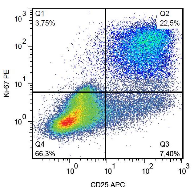
ARG55041 anti-Ki-67 antibody [Ki-67] (PE) FACS image
Flow Cytometry: Human peripheral blood mononuclear cells stimulated with PHA. Surface staining of ARG53800 anti-CD25 antibody [MEM-181] (APC) was followed by permeabilization and nuclear staining of ARG55041 anti-Ki-67 antibody [Ki-67] (PE).