ARG56320
anti-KHDRBS1 antibody
anti-KHDRBS1 antibody for ICC/IF,IHC-Formalin-fixed paraffin-embedded sections,Western blot and Human,Mouse,Rat
Overview
| Product Description | Rabbit Polyclonal antibody recognizes KHDRBS1 |
|---|---|
| Tested Reactivity | Hu, Ms, Rat |
| Tested Application | ICC/IF, IHC-P, WB |
| Host | Rabbit |
| Clonality | Polyclonal |
| Isotype | IgG |
| Target Name | KHDRBS1 |
| Antigen Species | Human |
| Immunogen | Synthetic peptide of Human KHDRBS1 |
| Conjugation | Un-conjugated |
| Alternate Names | KH domain-containing, RNA-binding, signal transduction-associated protein 1; p62; p68; p21 Ras GTPase-activating protein-associated p62; GAP-associated tyrosine phosphoprotein p62; Src-associated in mitosis 68 kDa protein; Sam68 |
Application Instructions
| Application Suggestion |
|
||||||||
|---|---|---|---|---|---|---|---|---|---|
| Application Note | * The dilutions indicate recommended starting dilutions and the optimal dilutions or concentrations should be determined by the scientist. | ||||||||
| Positive Control | MCF7 |
Properties
| Form | Liquid |
|---|---|
| Purification | Affinity purification with immunogen. |
| Buffer | PBS (pH 7.3), 0.02% Sodium azide and 50% Glycerol. |
| Preservative | 0.02% sodium azide |
| Stabilizer | 50% Glycerol |
| Storage Instruction | For continuous use, store undiluted antibody at 2-8°C for up to a week. For long-term storage, aliquot and store at -20°C. Storage in frost free freezers is not recommended. Avoid repeated freeze/thaw cycles. Suggest spin the vial prior to opening. The antibody solution should be gently mixed before use. |
| Note | For laboratory research only, not for drug, diagnostic or other use. |
Bioinformation
| Database Links | |
|---|---|
| Gene Symbol | KHDRBS1 |
| Gene Full Name | KH domain containing, RNA binding, signal transduction associated 1 |
| Background | This gene encodes a member of the K homology domain-containing, RNA-binding, signal transduction-associated protein family. The encoded protein appears to have many functions and may be involved in a variety of cellular processes, including alternative splicing, cell cycle regulation, RNA 3'-end formation, tumorigenesis, and regulation of human immunodeficiency virus gene expression. Alternative splicing results in multiple transcript variants. [provided by RefSeq, Dec 2012] |
| Function | Recruited and tyrosine phosphorylated by several receptor systems, for example the T-cell, leptin and insulin receptors. Once phosphorylated, functions as an adapter protein in signal transduction cascades by binding to SH2 and SH3 domain-containing proteins. Role in G2-M progression in the cell cycle. Represses CBP-dependent transcriptional activation apparently by competing with other nuclear factors for binding to CBP. Also acts as a putative regulator of mRNA stability and/or translation rates and mediates mRNA nuclear export. Positively regulates the association of constitutive transport element (CTE)-containing mRNA with large polyribosomes and translation initiation. According to some authors, is not involved in the nucleocytoplasmic export of unspliced (CTE)-containing RNA species according to. Isoform 3, which is expressed in growth-arrested cells only, inhibits S phase. [UniProt] |
| Calculated MW | 48 kDa |
| PTM | Tyrosine phosphorylated by several non-receptor tyrosine kinases, LCK, FYN and JAK3. Negatively correlates with ability to bind RNA but required for many interactions with proteins. Phosphorylation by PTK6 negatively regulates its RNA binding ability. Phosphorylation by PTK6 at Tyr-440 dictates the nulear localization of KHDRBS1. Phosphorylation at Tyr-387 disrupts interaction with APC. Phosphorylation at tyrosine residues by FYN inverts activity on modulation of BCL2L1 alternative splicing. Acetylated. Positively correlates with ability to bind RNA. Arginine methylation is required for nuclear localization. Also can affect interaction with other proteins. Inhibits interaction with Src-like SH3 domains, but not interaction with WW domains of WBP4/FBP21 AND FNBP4/FBP30. |
Images (2) Click the Picture to Zoom In
-
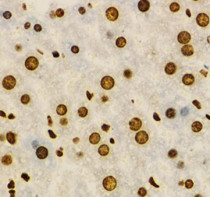
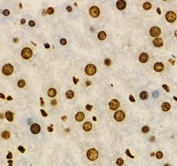
ARG56320 anti-KHDRBS1 antibody IHC-P image
Immunohistochemistry: Paraffin-embedded Mouse liver stained with ARG56320 anti-KHDRBS1 antibody at 1:100 dilution.
-
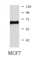
ARG56320 anti-KHDRBS1 antibody WB image
Western blot: MCF7 cell lysate stained with ARG56320 anti-KHDRBS1 antibody.