ARG55235
anti-ITPA antibody
anti-ITPA antibody for Flow cytometry,ICC/IF,Immunohistochemistry,Western blot and Human,Mouse,Rat
Metabolism antibody; Signaling Transduction antibody
Overview
| Product Description | Rabbit Polyclonal antibody recognizes ITPA |
|---|---|
| Tested Reactivity | Hu, Ms, Rat |
| Predict Reactivity | Bov, Chk, Xenopus |
| Tested Application | FACS, ICC/IF, IHC, WB |
| Host | Rabbit |
| Clonality | Polyclonal |
| Isotype | IgG |
| Target Name | ITPA |
| Antigen Species | Human |
| Immunogen | KLH-conjugated synthetic peptide corresponding to aa. 24-51 (N-terminus) of Human ITPA. |
| Conjugation | Un-conjugated |
| Alternate Names | Inosine triphosphatase; My049; EC 3.6.1.19; Inosine triphosphate pyrophosphatase; Non-canonical purine NTP pyrophosphatase; NTPase; C20orf37; Putative oncogene protein hlc14-06-p; ITPase; HLC14-06-P; Nucleoside-triphosphate diphosphatase; Nucleoside-triphosphate pyrophosphatase; dJ794I6.3; Non-standard purine NTP pyrophosphatase |
Application Instructions
| Application Suggestion |
|
||||||||||
|---|---|---|---|---|---|---|---|---|---|---|---|
| Application Note | * The dilutions indicate recommended starting dilutions and the optimal dilutions or concentrations should be determined by the scientist. |
Properties
| Form | Liquid |
|---|---|
| Purification | Purification with Protein A and immunogen peptide. |
| Buffer | PBS and 0.09% (W/V) Sodium azide |
| Preservative | 0.09% (W/V) Sodium azide |
| Storage Instruction | For continuous use, store undiluted antibody at 2-8°C for up to a week. For long-term storage, aliquot and store at -20°C or below. Storage in frost free freezers is not recommended. Avoid repeated freeze/thaw cycles. Suggest spin the vial prior to opening. The antibody solution should be gently mixed before use. |
| Note | For laboratory research only, not for drug, diagnostic or other use. |
Bioinformation
| Database Links | |
|---|---|
| Gene Symbol | ITPA |
| Gene Full Name | inosine triphosphatase (nucleoside triphosphate pyrophosphatase) |
| Background | This gene encodes an inosine triphosphate pyrophosphohydrolase. The encoded protein hydrolyzes inosine triphosphate and deoxyinosine triphosphate to the monophosphate nucleotide and diphosphate. This protein, which is a member of the HAM1 NTPase protein family, is found in the cytoplasm and acts as a homodimer. Defects in the encoded protein can result in inosine triphosphate pyrophosphorylase deficiency which causes an accumulation of ITP in red blood cells. Alternate splicing results in multiple transcript variants. [provided by RefSeq, Jun 2012] |
| Function | Pyrophosphatase that hydrolyzes the non-canonical purine nucleotides inosine triphosphate (ITP), deoxyinosine triphosphate (dITP) as well as 2'-deoxy-N-6-hydroxylaminopurine triposphate (dHAPTP) and xanthosine 5'-triphosphate (XTP) to their respective monophosphate derivatives. The enzyme does not distinguish between the deoxy- and ribose forms. Probably excludes non-canonical purines from RNA and DNA precursor pools, thus preventing their incorporation into RNA and DNA and avoiding chromosomal lesions. [UniProt] |
| Cellular Localization | Cytoplasm {ECO:0000255|HAMAP-Rule:MF_03148, ECO:0000269|PubMed:11278832} |
| Research Area | Metabolism antibody; Signaling Transduction antibody |
| Calculated MW | 21 kDa |
Images (4) Click the Picture to Zoom In
-
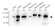
ARG55235 anti-ITPA antibody WB image
Western blot: 30 μg of HeLa, Jurkat, Mouse brain, Mouse liver, Mouse kidney, Rat brain and Rat liver lysates stained with ARG55235 anti-ITPA antibody at 1:500 dilution.
-
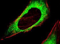
ARG55235 anti-ITPA antibody ICC/IF image
Immunofluorescence: HeLa cells stained with ARG55235 anti-ITPA antibody (green) at 1:25 dilution. Cytoplasmic actin was counterstained with Dylight Fluor® 554 conjugated Phalloidin (red).
-
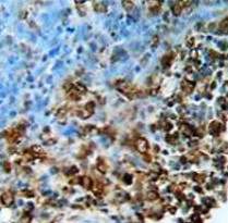
ARG55235 anti-ITPA antibody IHC-P image
Immunohistochemistry: Formalin-fixed and paraffin-embedded Human lung carcinoma stained with ARG55235 anti-ITPA antibody.
-
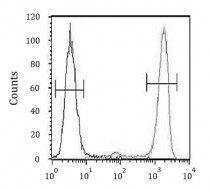
ARG55235 anti-ITPA antibody FACS image
Flow Cytometry: A375 cells stained with ARG55235 anti-ITPA antibody (right histogram) or without primary antibody control (left histogram), followed by incubation with FITC labelled secondary antibody.


