ARG63136
anti-ITK antibody
anti-ITK antibody for Flow cytometry,ICC/IF,Western blot and Human
Signaling Transduction antibody
Overview
| Product Description | Goat Polyclonal antibody recognizes ITK |
|---|---|
| Tested Reactivity | Hu |
| Predict Reactivity | Ms |
| Tested Application | FACS, ICC/IF, WB |
| Host | Goat |
| Clonality | Polyclonal |
| Isotype | IgG |
| Target Name | ITK |
| Antigen Species | Human |
| Immunogen | C-RLLRQLAEIAESGL |
| Conjugation | Un-conjugated |
| Alternate Names | LPFS1; Interleukin-2-inducible T-cell kinase; T-cell-specific kinase; Tyrosine-protein kinase ITK/TSK; PSCTK2; EMT; Kinase EMT; Tyrosine-protein kinase Lyk; LYK; IL-2-inducible T-cell kinase; EC 2.7.10.2 |
Application Instructions
| Application Suggestion |
|
||||||||
|---|---|---|---|---|---|---|---|---|---|
| Application Note | WB: Recommend incubate at RT for 1h. * The dilutions indicate recommended starting dilutions and the optimal dilutions or concentrations should be determined by the scientist. |
Properties
| Form | Liquid |
|---|---|
| Purification | Purified from goat serum by antigen affinity chromatography. |
| Buffer | Tris saline (pH 7.3), 0.02% Sodium azide and 0.5% BSA. |
| Preservative | 0.02% Sodium azide |
| Stabilizer | 0.5% BSA |
| Concentration | 0.5 mg/ml |
| Storage Instruction | For continuous use, store undiluted antibody at 2-8°C for up to a week. For long-term storage, aliquot and store at -20°C or below. Storage in frost free freezers is not recommended. Avoid repeated freeze/thaw cycles. Suggest spin the vial prior to opening. The antibody solution should be gently mixed before use. |
| Note | For laboratory research only, not for drug, diagnostic or other use. |
Bioinformation
| Database Links | |
|---|---|
| Background | This gene encodes an intracellular tyrosine kinase expressed in T-cells. The protein contains both SH2 and SH3 domains which are often found in intracellular kinases. It is thought to play a role in T-cell proliferation and differentiation. [provided by RefSeq, Jul 2008] |
| Research Area | Signaling Transduction antibody |
| Calculated MW | 72 kDa |
| PTM | Phosphorylated at Tyr-512 in the activation loop of the kinase domain by LCK. Subsequent autophosphorylation at Tyr-180 leads to the kinase activation. The autophosphorylated Tyr-180 lies within the substrate binding sequence of the SH3 domain. Ubiquitinated. |
Images (3) Click the Picture to Zoom In
-

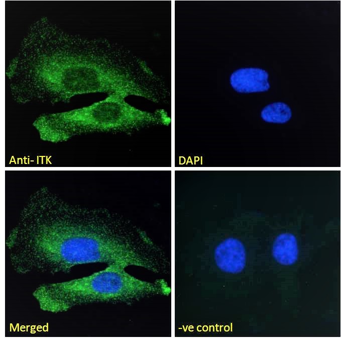
ARG63136 anti-ITK antibody ICC/IF image
Immunofluorescence: Paraformaldehyde fixed HeLa cells permeabilized with 0.15% Triton. Cells were stained with ARG63136 anti-ITK antibody (green) at 10 µg/ml dilution for 1 hour. DAPI (blue) for nuclear staining. Negative control: Unimmunized goat IgG (green) at 10 µg/ml dilution.
-

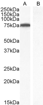
ARG63136 anti-ITK antibody WB image
Western blot: 35 µg of Jurkat nuclear lysate (A) and Human parathyroid gland (B, negative control) lysate (in RIPA buffer) stained with ARG63136 anti-ITK antibody at 1 µg/ml dilution and incubated at RT for 1 hour.
-

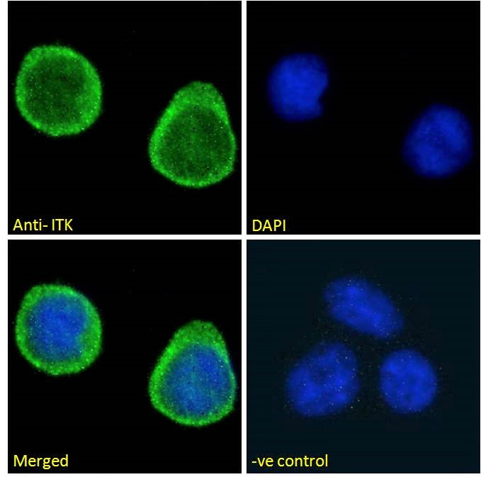
ARG63136 anti-ITK antibody ICC/IF image
Immunofluorescence: Paraformaldehyde fixed Jurkat cells permeabilized with 0.15% Triton. Cells were stained with ARG63136 anti-ITK antibody (green) at 10 µg/ml dilution for 1 hour. DAPI (blue) for nuclear staining. Negative control: Unimmunized goat IgG (green) at 10 µg/ml dilution.