ARG41456
anti-ITCH / AIF4 antibody
anti-ITCH / AIF4 antibody for Western blot and Human,Mouse,Rat
Overview
| Product Description | Rabbit Polyclonal antibody recognizes ITCH / AIF4 |
|---|---|
| Tested Reactivity | Hu, Ms, Rat |
| Tested Application | WB |
| Host | Rabbit |
| Clonality | Polyclonal |
| Isotype | IgG |
| Target Name | ITCH / AIF4 |
| Antigen Species | Human |
| Immunogen | Synthetic peptide of Human ITCH / AIF4. |
| Conjugation | Un-conjugated |
| Alternate Names | EC 6.3.2.-; Itch; Atrophin-1-interacting protein 4; ADMFD; E3 ubiquitin-protein ligase Itchy homolog; NFE2-associated polypeptide 1; AIF4; AIP4; NAPP1 |
Application Instructions
| Application Suggestion |
|
||||
|---|---|---|---|---|---|
| Application Note | * The dilutions indicate recommended starting dilutions and the optimal dilutions or concentrations should be determined by the scientist. | ||||
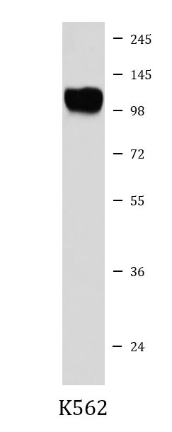
| Positive Control | K562 | ||||
| Observed Size | ~ 105 kDa |
Properties
| Form | Liquid |
|---|---|
| Purification | Affinity purified. |
| Buffer | PBS (pH 7.4), 150 mM NaCl, 0.02% Sodium azide and 50% Glycerol. |
| Preservative | 0.02% Sodium azide |
| Stabilizer | 50% Glycerol |
| Storage Instruction | For continuous use, store undiluted antibody at 2-8°C for up to a week. For long-term storage, aliquot and store at -20°C. Storage in frost free freezers is not recommended. Avoid repeated freeze/thaw cycles. Suggest spin the vial prior to opening. The antibody solution should be gently mixed before use. |
| Note | For laboratory research only, not for drug, diagnostic or other use. |
Bioinformation
| Database Links |
Swiss-port # Q8C863 Mouse E3 ubiquitin-protein ligase Itchy Swiss-port # Q96J02 Human E3 ubiquitin-protein ligase Itchy homolog |
|---|---|
| Gene Symbol | ITCH |
| Gene Full Name | itchy E3 ubiquitin protein ligase |
| Background | This gene encodes a member of the Nedd4 family of HECT domain E3 ubiquitin ligases. HECT domain E3 ubiquitin ligases transfer ubiquitin from E2 ubiquitin-conjugating enzymes to protein substrates, thus targeting specific proteins for lysosomal degradation. The encoded protein plays a role in multiple cellular processes including erythroid and lymphoid cell differentiation and the regulation of immune responses. Mutations in this gene are a cause of syndromic multisystem autoimmune disease. Alternatively spliced transcript variants encoding multiple isoforms have been observed for this gene. [provided by RefSeq, Mar 2012] |
| Function | Acts as an E3 ubiquitin-protein ligase which accepts ubiquitin from an E2 ubiquitin-conjugating enzyme in the form of a thioester and then directly transfers the ubiquitin to targeted substrates. It catalyzes 'Lys-29'-, 'Lys-48'- and 'Lys-63'-linked ubiquitin conjugation. It is involved in the control of inflammatory signaling pathways. Is an essential component of a ubiquitin-editing protein complex, comprising also TNFAIP3, TAX1BP1 and RNF11, that ensures the transient nature of inflammatory signaling pathways. Promotes the association of the complex after TNF stimulation. Once the complex is formed, TNFAIP3 deubiquitinates 'Lys-63' polyubiquitin chains on RIPK1 and catalyzes the formation of 'Lys-48'-polyubiquitin chains. This leads to RIPK1 proteasomal degradation and consequently termination of the TNF- or LPS-mediated activation of NFKB1. Ubiquitinates RIPK2 by 'Lys-63'-linked conjugation and influences NOD2-dependent signal transduction pathways. Regulates the transcriptional activity of several transcription factors, and probably plays an important role in the regulation of immune response. Ubiquitinates NFE2 by 'Lys-63' linkages and is implicated in the control of the development of hematopoietic lineages. Critical regulator of T-helper (TH2) cytokine development through its ability to induce JUNB ubiquitination and degradation (By similarity). Ubiquitinates SNX9. Ubiquitinates CXCR4 and HGS/HRS and regulates sorting of CXCR4 to the degradative pathway. It is involved in the negative regulation of MAVS-dependent cellular antiviral responses. Ubiquitinates MAVS through 'Lys-48'-linked conjugation resulting in MAVS proteasomal degradation. Involved in the regulation of apoptosis and reactive oxygen species levels through the ubiquitination and proteasomal degradation of TXNIP. Mediates the antiapoptotic activity of epidermal growth factor through the ubiquitination and proteasomal degradation of p15 BID. Targets DTX1 for lysosomal degradation and controls NOTCH1 degradation, in the absence of ligand, through 'Lys-29'-linked polyubiquitination. Ubiquitinates BRAT1 and this ubiquitination is enhanced in the presence of NDFIP1. [UniProt] |
| Cellular Localization | Cell membrane; Peripheral membrane protein; Cytoplasmic side. Cytoplasm. Nucleus. Early endosome membrane; Peripheral membrane protein; Cytoplasmic side. Endosome membrane; Peripheral membrane protein; Cytoplasmic side. Note=May be recruited to exosomes by NDFIP1. Localizes to plasma membrane upon CXCL12 stimulation where it co-localizes with CXCL4. Localization to early endosomes is increased upon CXCL12 stimulation where it co-localizes with DTX3L and CXCL4. [UniProt] |
| Calculated MW | 103 kDa |
| PTM | On T-cell activation, phosphorylation by the JNK cascade on serine and threonine residues surrounding the PRR domain accelerates the ubiquitination and degradation of JUN and JUNB. The increased ITCH catalytic activity due to phosphorylation by JNK1 may occur due to a conformational change disrupting the interaction between the PRR/WW motifs domain and the HECT domain and, thus exposing the HECT domain (By similarity). Phosphorylation by FYN reduces interaction with JUNB and negatively controls JUN ubiquitination and degradation. Ubiquitinated; autopolyubiquitination with 'Lys-63' linkages which does not lead to protein degradation. [UniProt] |
Images (1) Click the Picture to Zoom In
-
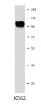
ARG41456 anti-ITCH / AIF4 antibody WB image
Western blot: K562 cell lysate stained with ARG41456 anti-ITCH / AIF4 antibody.