ARG66967
anti-IRE1a antibody
anti-IRE1a antibody for ICC/IF,IHC-Formalin-fixed paraffin-embedded sections,Western blot and Human,Mouse
Cell Biology and Cellular Response antibody; Cell Death antibody; Gene Regulation antibody; Signaling Transduction antibody
Overview
| Product Description | Rabbit Polyclonal antibody recognizes IRE1a |
|---|---|
| Tested Reactivity | Hu, Ms |
| Predict Reactivity | Rat |
| Tested Application | ICC/IF, IHC-P, WB |
| Host | Rabbit |
| Clonality | Polyclonal |
| Isotype | IgG |
| Target Name | IRE1a |
| Antigen Species | Human |
| Immunogen | Synthetic peptide around the middle region of Human IRE1a protein. |
| Conjugation | Un-conjugated |
| Alternate Names | Ire1-alpha; Serine/threonine-protein kinase/endoribonuclease IRE1; IRE1a; Endoplasmic reticulum-to-nucleus signaling 1; EC 3.1.26.-; IRE1; Inositol-requiring protein 1; IRE1P; EC 2.7.11.1; hIRE1p |
Application Instructions
| Application Suggestion |
|
||||||||
|---|---|---|---|---|---|---|---|---|---|
| Application Note | * The dilutions indicate recommended starting dilutions and the optimal dilutions or concentrations should be determined by the scientist. | ||||||||
| Observed Size | ~130 kDa |
Properties
| Form | Liquid |
|---|---|
| Purification | Affinity purified. |
| Buffer | 100 mM Tris Glycine (pH 7.0), 0.025% ProClin 300 and 20% Glycerol. |
| Preservative | 0.025% ProClin 300 |
| Stabilizer | 20% Glycerol |
| Storage Instruction | For continuous use, store undiluted antibody at 2-8°C for up to a week. For long-term storage, aliquot and store at -20°C or below. Storage in frost free freezers is not recommended. Avoid repeated freeze/thaw cycles. Suggest spin the vial prior to opening. The antibody solution should be gently mixed before use. |
| Note | For laboratory research only, not for drug, diagnostic or other use. |
Bioinformation
| Database Links |
Swiss-port # O75460 Human Serine/threonine-protein kinase/endoribonuclease IRE1 Swiss-port # Q9EQY0 Mouse Serine/threonine-protein kinase/endoribonuclease IRE1 |
|---|---|
| Gene Symbol | ERN1 |
| Gene Full Name | endoplasmic reticulum to nucleus signaling 1 |
| Background | IRE1p Antibody: Accumulation of malfolded proteins in the endoplasmic reticulum (ER) activates the unfolded protein response (UPR) and the upregulation of the ER molecular chaperones GRP78 and GRP 94. These proteins are normally bound to ER transmembrane proteins such as IRE1p and ATF6 but ER stress causes their dissociation. This allows IRE1p, a serine-threonine protein kinase to transduce the unfolded protein signal from the ER to the nucleus. IRE1p also has an endoribonuclease activity that is required to splice X-box binding protein (XBP1) mRNA converting it to a potent UPR transcriptional activation. Depletion of IRE1p through the expression of a dominant negative form of IRE1p has no effect on transfected cells, but cell death via apoptosis occurs under stress conditions that cause unfolded proteins to accumulate in the ER. Two alternatively spliced transcript variants encoding different isoforms have been found for this gene. |
| Function | Senses unfolded proteins in the lumen of the endoplasmic reticulum via its N-terminal domain which leads to enzyme auto-activation. The active endoribonuclease domain splices XBP1 mRNA to generate a new C-terminus, converting it into a potent unfolded-protein response transcriptional activator and triggering growth arrest and apoptosis. [UniProt] |
| Research Area | Cell Biology and Cellular Response antibody; Cell Death antibody; Gene Regulation antibody; Signaling Transduction antibody |
| Calculated MW | 110 kDa |
| PTM | Autophosphorylated. ADP-ribosylated by PARP16 upon ER stress, which increases both kinase and endonuclease activities. |
Images (3) Click the Picture to Zoom In
-
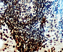
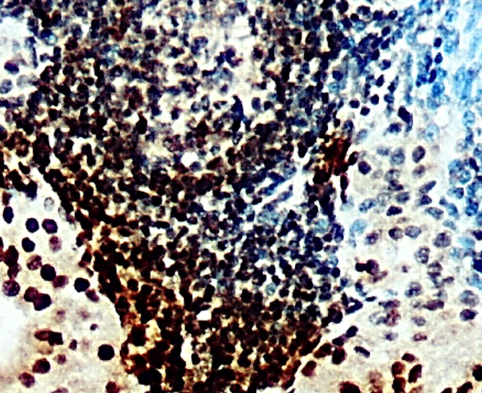
ARG66967 anti-IRE1a antibody IHC-P image
Immunohistochemistry: Paraffin-embedded Human Tonsil cancer tissue stained with ARG66967 anti-IRE1a antibody at 1:100 dilution.
-
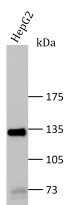
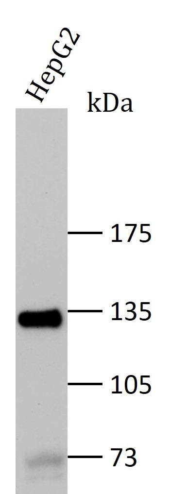
ARG66967 anti-IRE1a antibody WB image
Western blot: Lysate from HepG2 treated with thapsigargin (TG) or not, stained with ARG66967 anti-IRE1a antibody at 1:500 dilution, overnight at 4°C.
-
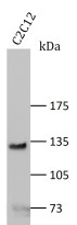
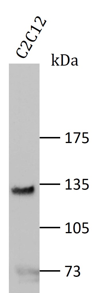
ARG66967 anti-IRE1a antibody WB image
Western blot: C2C12 cell lysate stained with ARG66967 anti-IRE1a antibody at 1:500 dilution, overnight at 4°C.