ARG59050
anti-INPPL1 antibody
anti-INPPL1 antibody for IHC-Formalin-fixed paraffin-embedded sections and Human,Mouse,Rat
Overview
| Product Description | Rabbit Polyclonal antibody recognizes INPPL1 |
|---|---|
| Tested Reactivity | Hu, Ms, Rat |
| Tested Application | IHC-P |
| Host | Rabbit |
| Clonality | Polyclonal |
| Isotype | IgG |
| Target Name | INPPL1 |
| Antigen Species | Human |
| Immunogen | Recombinant protein corresponding to R1172-K1258 of Human INPPL1. |
| Conjugation | Un-conjugated |
| Alternate Names | INPPL-1; Protein 51C; Inositol polyphosphate phosphatase-like protein 1; OPSMD; SHIP2; EC 3.1.3.86; SHIP-2; SH2 domain-containing inositol 5'-phosphatase 2; SH2 domain-containing inositol phosphatase 2; Phosphatidylinositol 3,4,5-trisphosphate 5-phosphatase 2 |
Application Instructions
| Application Suggestion |
|
||||
|---|---|---|---|---|---|
| Application Note | IHC-P: Antigen Retrieval: Heat mediated was performed in Citrate buffer (pH 6.0) for 20 min. * The dilutions indicate recommended starting dilutions and the optimal dilutions or concentrations should be determined by the scientist. |
||||
| Observed Size | ~ 140 - 160 kDa |
Properties
| Form | Liquid |
|---|---|
| Purification | Affinity purification with immunogen. |
| Buffer | 0.2% Na2HPO4, 0.9% NaCl, 0.05% Sodium azide and 4% Trehalose. |
| Preservative | 0.05% Sodium azide |
| Stabilizer | 4% Trehalose |
| Concentration | 0.5 mg/ml |
| Storage Instruction | For continuous use, store undiluted antibody at 2-8°C for up to a week. For long-term storage, aliquot and store at -20°C or below. Storage in frost free freezers is not recommended. Avoid repeated freeze/thaw cycles. Suggest spin the vial prior to opening. The antibody solution should be gently mixed before use. |
| Note | For laboratory research only, not for drug, diagnostic or other use. |
Bioinformation
| Database Links | |
|---|---|
| Gene Symbol | INPPL1 |
| Gene Full Name | inositol polyphosphate phosphatase-like 1 |
| Background | The protein encoded by this gene is an SH2-containing 5'-inositol phosphatase that is involved in the regulation of insulin function. The encoded protein also plays a role in the regulation of epidermal growth factor receptor turnover and actin remodelling. Additionally, this gene supports metastatic growth in breast cancer and is a valuable biomarker for breast cancer. [provided by RefSeq, Jan 2009] |
| Function | Phosphatidylinositol (PtdIns) phosphatase that specifically hydrolyzes the 5-phosphate of phosphatidylinositol-3,4,5-trisphosphate (PtdIns(3,4,5)P3) to produce PtdIns(3,4)P2, thereby negatively regulating the PI3K (phosphoinositide 3-kinase) pathways. Plays a central role in regulation of PI3K-dependent insulin signaling, although the precise molecular mechanisms and signaling pathways remain unclear. While overexpression reduces both insulin-stimulated MAP kinase and Akt activation, its absence does not affect insulin signaling or GLUT4 trafficking. Confers resistance to dietary obesity. May act by regulating AKT2, but not AKT1, phosphorylation at the plasma membrane. Part of a signaling pathway that regulates actin cytoskeleton remodeling. Required for the maintenance and dynamic remodeling of actin structures as well as in endocytosis, having a major impact on ligand-induced EGFR internalization and degradation. Participates in regulation of cortical and submembraneous actin by hydrolyzing PtdIns(3,4,5)P3 thereby regulating membrane ruffling. Regulates cell adhesion and cell spreading. Required for HGF-mediated lamellipodium formation, cell scattering and spreading. Acts as a negative regulator of EPHA2 receptor endocytosis by inhibiting via PI3K-dependent Rac1 activation. Acts as a regulator of neuritogenesis by regulating PtdIns(3,4,5)P3 level and is required to form an initial protrusive pattern, and later, maintain proper neurite outgrowth. Acts as a negative regulator of the FC-gamma-RIIA receptor (FCGR2A). Mediates signaling from the FC-gamma-RIIB receptor (FCGR2B), playing a central role in terminating signal transduction from activating immune/hematopoietic cell receptor systems. Involved in EGF signaling pathway. Upon stimulation by EGF, it is recruited by EGFR and dephosphorylates PtdIns(3,4,5)P3. Plays a negative role in regulating the PI3K-PKB pathway, possibly by inhibiting PKB activity. Down-regulates Fc-gamma-R-mediated phagocytosis in macrophages independently of INPP5D/SHIP1. In macrophages, down-regulates NF-kappa-B-dependent gene transcription by regulating macrophage colony-stimulating factor (M-CSF)-induced signaling. May also hydrolyze PtdIns(1,3,4,5)P4, and could thus affect the levels of the higher inositol polyphosphates like InsP6. Involved in endochondral ossification. [UniProt] |
| Cellular Localization | Cytoplasm, cytosol. Cytoplasm, cytoskeleton. Membrane; Peripheral membrane protein. Cell projection, filopodium. Cell projection, lamellipodium. [UniProt] |
| Calculated MW | 139 kDa |
| PTM | Tyrosine phosphorylated by the members of the SRC family after exposure to a diverse array of extracellular stimuli such as insulin, growth factors such as EGF or PDGF, chemokines, integrin ligands and hypertonic and oxidative stress. May be phosphorylated upon IgG receptor FCGR2B-binding. Phosphorylated at Tyr-986 following cell attachment and spreading. Phosphorylated at Tyr-1162 following EGF signaling pathway stimulation. Phosphorylated at Thr-958 in response to PDGF. [UniProt] |
Images (7) Click the Picture to Zoom In
-
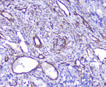
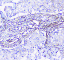
ARG59050 anti-INPPL1 antibody IHC-P image
Immunohistochemistry: Paraffin-embedded Human gastric cancer tissue. Antigen Retrieval: Heat mediated was performed in Citrate buffer (pH 6.0, epitope retrieval solution) for 20 min. The tissue section was blocked with 10% goat serum. The tissue section was then stained with ARG59050 anti-INPPL1 antibody at 1 µg/ml dilution, overnight at 4°C.
-
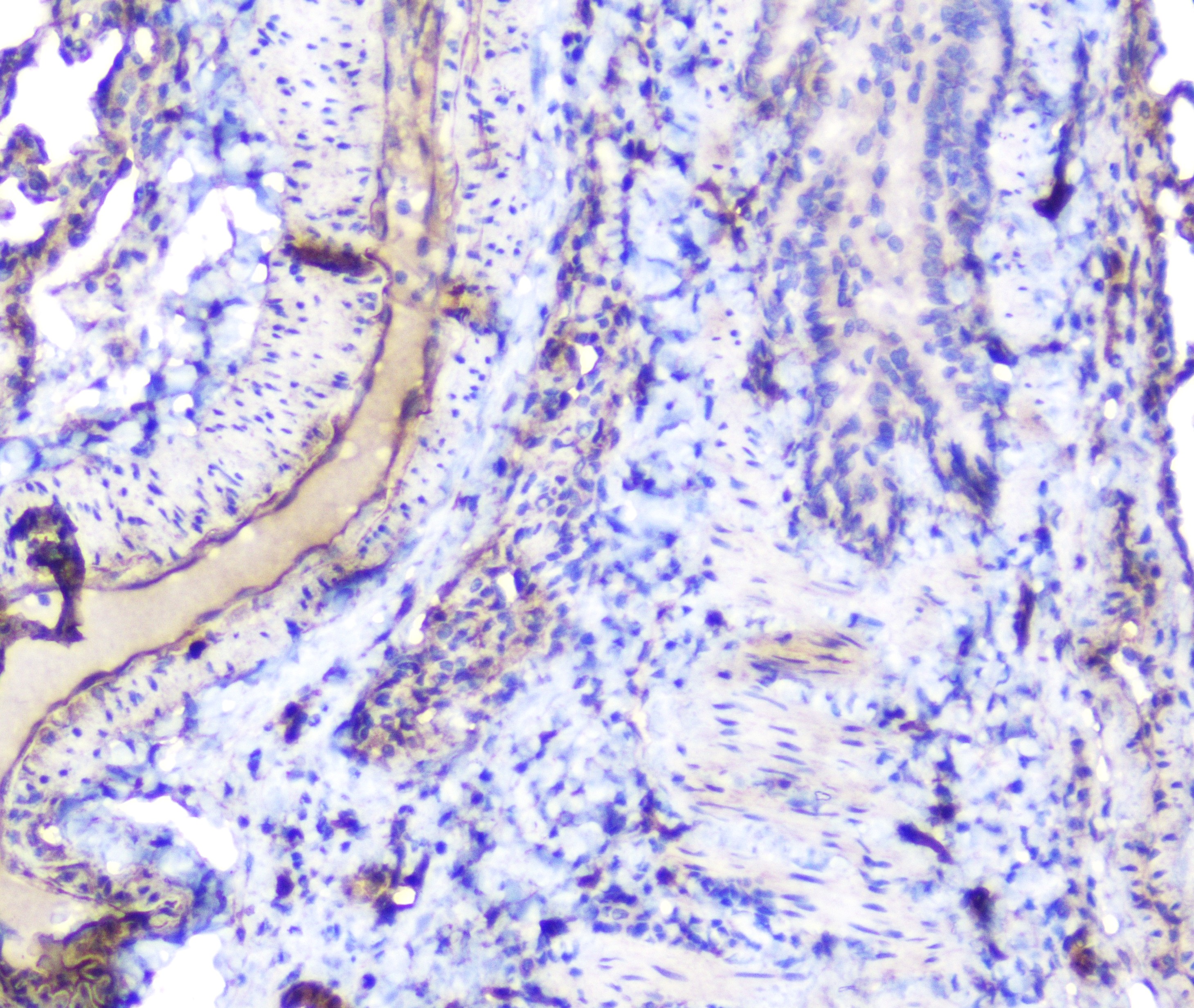
ARG59050 anti-INPPL1 antibody IHC-P image
Immunohistochemistry: Paraffin-embedded Human lung cancer tissue. Antigen Retrieval: Heat mediated was performed in Citrate buffer (pH 6.0, epitope retrieval solution) for 20 min. The tissue section was blocked with 10% goat serum. The tissue section was then stained with ARG59050 anti-INPPL1 antibody at 1 µg/ml dilution, overnight at 4°C.
-
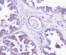
ARG59050 anti-INPPL1 antibody IHC-P image
Immunohistochemistry: Paraffin-embedded Human ovary cancer tissue. Antigen Retrieval: Heat mediated was performed in Citrate buffer (pH 6.0, epitope retrieval solution) for 20 min. The tissue section was blocked with 10% goat serum. The tissue section was then stained with ARG59050 anti-INPPL1 antibody at 1 µg/ml dilution, overnight at 4°C.
-
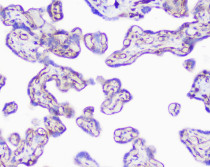
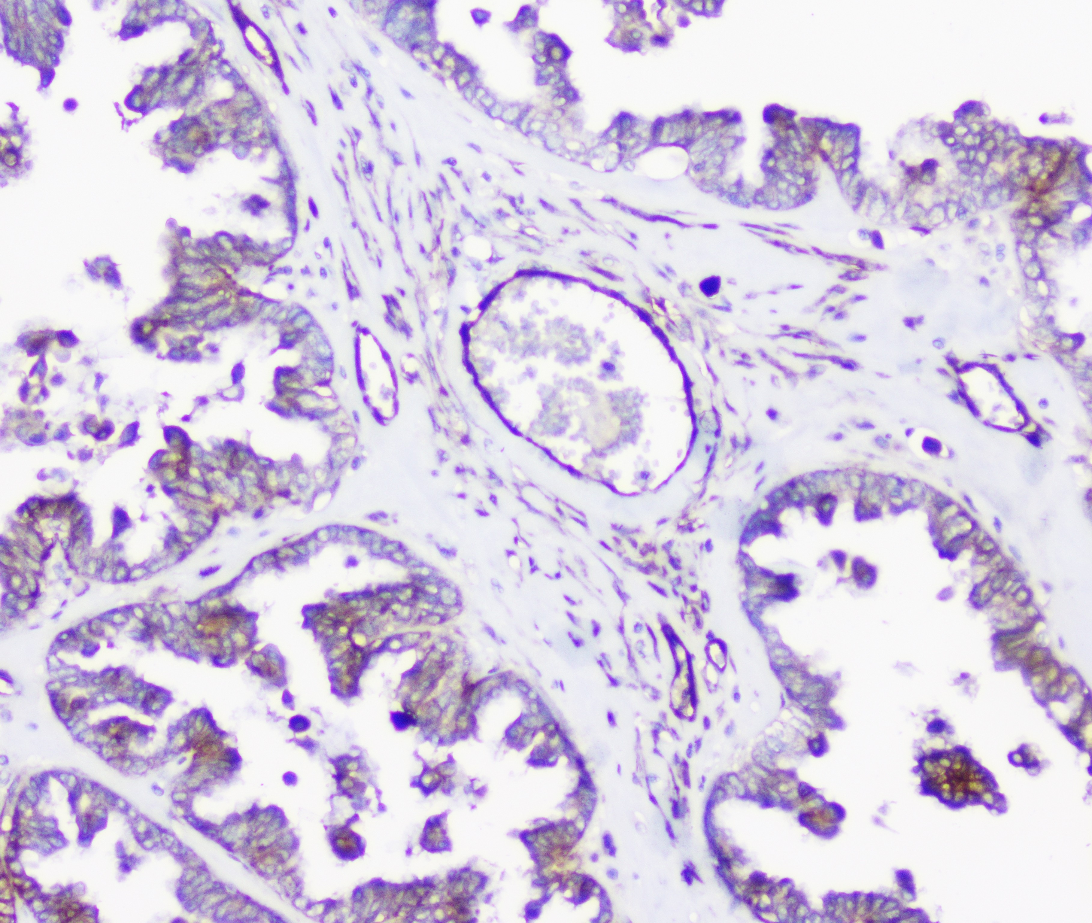
ARG59050 anti-INPPL1 antibody IHC-P image
Immunohistochemistry: Paraffin-embedded Human placenta tissue. Antigen Retrieval: Heat mediated was performed in Citrate buffer (pH 6.0, epitope retrieval solution) for 20 min. The tissue section was blocked with 10% goat serum. The tissue section was then stained with ARG59050 anti-INPPL1 antibody at 1 µg/ml dilution, overnight at 4°C.
-
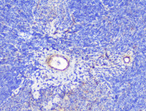
ARG59050 anti-INPPL1 antibody IHC-P image
Immunohistochemistry: Paraffin-embedded Mouse spleen tissue. Antigen Retrieval: Heat mediated was performed in Citrate buffer (pH 6.0, epitope retrieval solution) for 20 min. The tissue section was blocked with 10% goat serum. The tissue section was then stained with ARG59050 anti-INPPL1 antibody at 1 µg/ml dilution, overnight at 4°C.
-
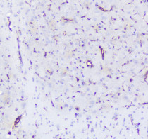
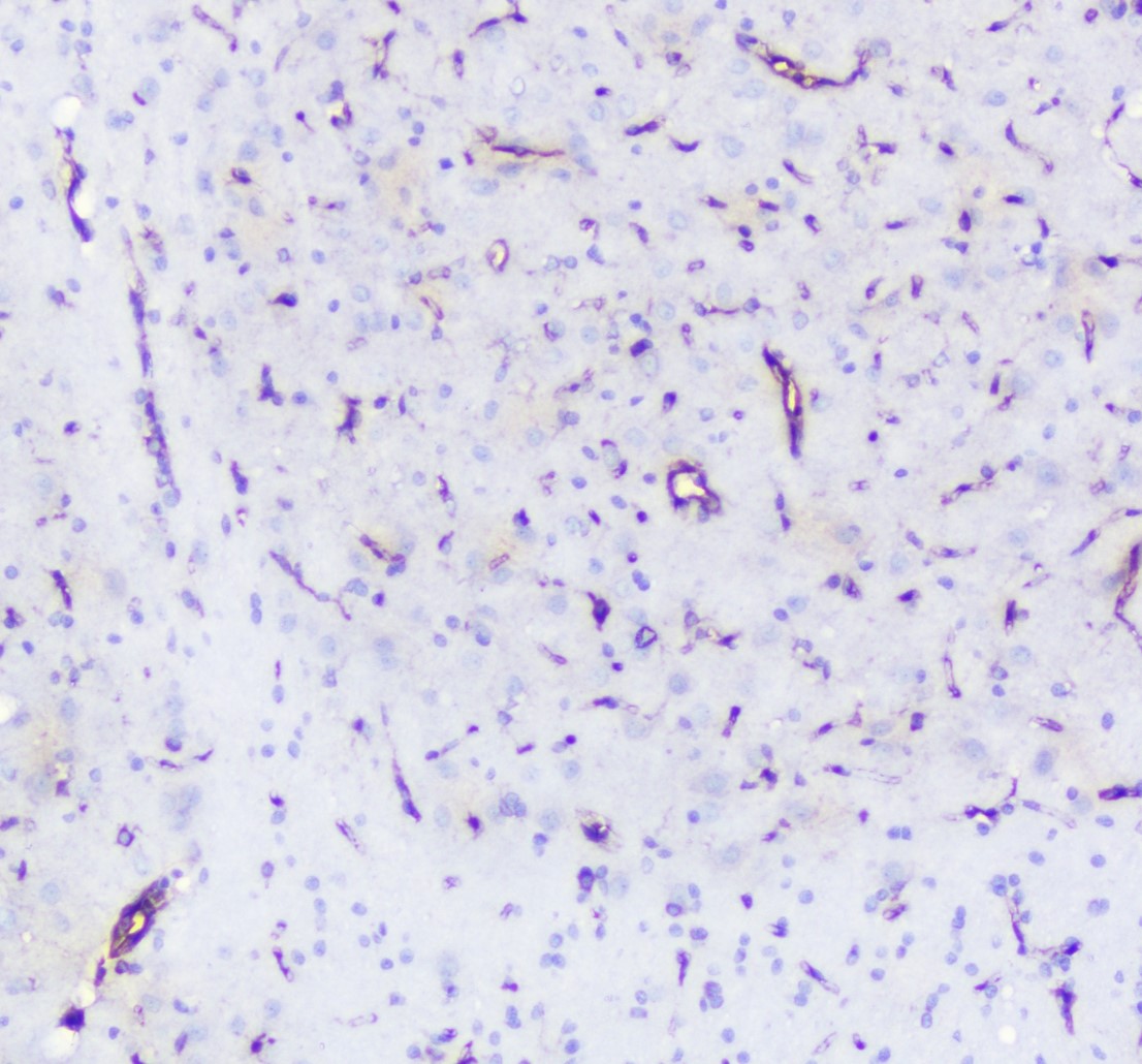
ARG59050 anti-INPPL1 antibody IHC-P image
Immunohistochemistry: Paraffin-embedded Rat brain tissue. Antigen Retrieval: Heat mediated was performed in Citrate buffer (pH 6.0, epitope retrieval solution) for 20 min. The tissue section was blocked with 10% goat serum. The tissue section was then stained with ARG59050 anti-INPPL1 antibody at 1 µg/ml dilution, overnight at 4°C.
-

ARG59050 anti-INPPL1 antibody IHC-P image
Immunohistochemistry: Paraffin-embedded Rat lung tissue. Antigen Retrieval: Heat mediated was performed in Citrate buffer (pH 6.0, epitope retrieval solution) for 20 min. The tissue section was blocked with 10% goat serum. The tissue section was then stained with ARG59050 anti-INPPL1 antibody at 1 µg/ml dilution, overnight at 4°C.