ARG66000
anti-IL5 antibody
anti-IL5 antibody for ELISA,IHC-Formalin-fixed paraffin-embedded sections,Neutralizing,Western blot and Human
Overview
| Product Description | Rabbit Polyclonal antibody recognizes IL5 |
|---|---|
| Tested Reactivity | Hu |
| Tested Application | ELISA, IHC-P, Neut, WB |
| Host | Rabbit |
| Clonality | Polyclonal |
| Isotype | IgG |
| Target Name | IL5 |
| Antigen Species | Human |
| Immunogen | E. coli derived recombinant Human IL5. (MIPTEIPTSA LVKETLALLS THRTLLIANE TLRIPVPVHK NHQLCTEEIF QGIGTLESQT VQGGTVERLF KNLSLIKKYI DGQKKKCGEE RRRVNQFLDY LQEFLGVMNT EWIIES) |
| Conjugation | Un-conjugated |
| Alternate Names | Eosinophil differentiation factor; EDF; IL-5; TRF; T-cell replacing factor; B-cell differentiation factor I; Interleukin-5 |
Application Instructions
| Application Suggestion |
|
||||||||||
|---|---|---|---|---|---|---|---|---|---|---|---|
| Application Note | * The dilutions indicate recommended starting dilutions and the optimal dilutions or concentrations should be determined by the scientist. |
Properties
| Form | Liquid |
|---|---|
| Purification | Affinity purification with immunogen. |
| Buffer | PBS (pH 7.2) |
| Concentration | 1 mg/ml |
| Storage Instruction | For continuous use, store undiluted antibody at 2-8°C for up to a week. For long-term storage, aliquot and store at -20°C or below. Storage in frost free freezers is not recommended. Avoid repeated freeze/thaw cycles. Suggest spin the vial prior to opening. The antibody solution should be gently mixed before use. |
| Note | For laboratory research only, not for drug, diagnostic or other use. |
Bioinformation
| Database Links | |
|---|---|
| Gene Symbol | IL5 |
| Gene Full Name | interleukin 5 |
| Background | This gene encodes a cytokine that acts as a growth and differentiation factor for both B cells and eosinophils. The encoded cytokine plays a major role in the regulation of eosinophil formation, maturation, recruitment and survival. The increased production of this cytokine may be related to pathogenesis of eosinophil-dependent inflammatory diseases. This cytokine functions by binding to its receptor, which is a heterodimer, whose beta subunit is shared with the receptors for interleukine 3 (IL3) and colony stimulating factor 2 (CSF2/GM-CSF). This gene is located on chromosome 5 within a cytokine gene cluster which includes interleukin 4 (IL4), interleukin 13 (IL13), and CSF2 . This gene, IL4, and IL13 may be regulated coordinately by long-range regulatory elements spread over 120 kilobases on chromosome 5q31. [provided by RefSeq, Jul 2013] |
| Function | Factor that induces terminal differentiation of late-developing B-cells to immunoglobulin secreting cells. [UniProt] |
| Calculated MW | 15 kDa |
Images (4) Click the Picture to Zoom In
-
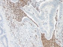
ARG66000 anti-IL5 antibody IHC-P image
Immunohistochemistry: Formalin-fixed and paraffin-embedded sections of Human colon and rectum adenocarcinoma. The recommended ARG66000 anti-IL5 antibody concentration is 0.25 µg/ml with an overnight incubation at 4°C. An HRP-labeled polymer detection system was used with a DAB chromogen.
-
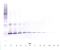
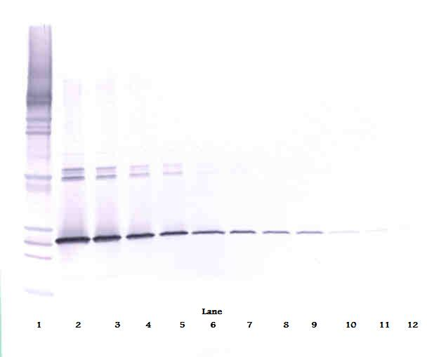
ARG66000 anti-IL5 antibody WB image
Western blot: 250 - 0.24 ng of Human IL-5 stained with ARG66000 anti-IL5 antibody, under non-reducing conditions.
-

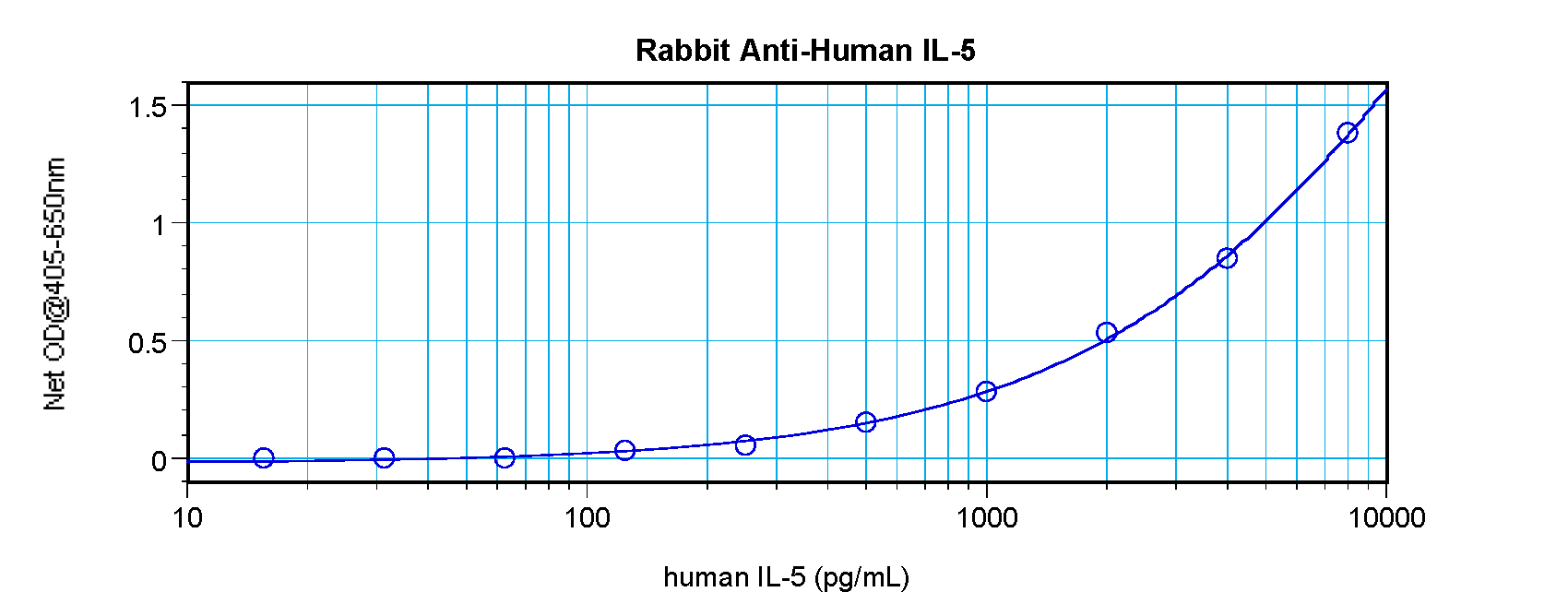
ARG66000 anti-IL5 antibody standard curve image
Sandwich ELISA: ARG66000 anti-IL5 antibody as a capture antibody at 0.5 - 2.0 µg/ml combined with ARG66001 anti-IL5 antibody (Biotin) as a detection antibody. Results of a typical standard run with optical density reading at 405 - 650 nm.
-
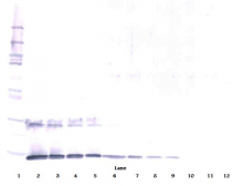
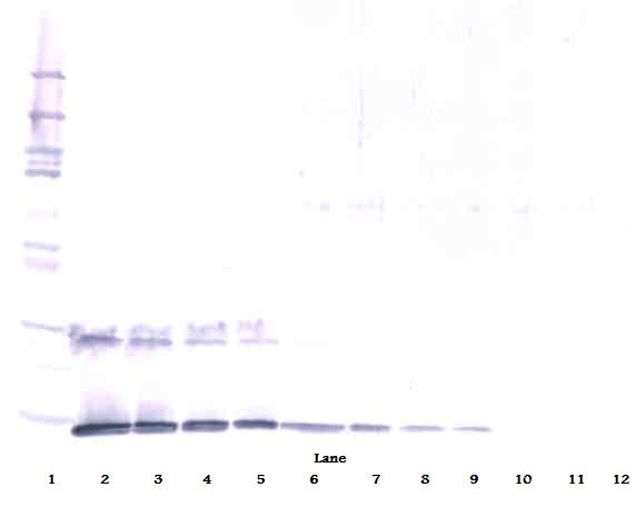
ARG66000 anti-IL5 antibody WB image
Western blot: 250 - 0.24 ng of Human IL-5 stained with ARG66000 anti-IL5 antibody, under reducing conditions.