ARG56952
anti-IL33 antibody [4E9]
anti-IL33 antibody [4E9] for Flow cytometry,ICC/IF,Western blot and Human
Overview
| Product Description | Mouse Monoclonal antibody [4E9] recognizes IL33 |
|---|---|
| Tested Reactivity | Hu |
| Tested Application | FACS, ICC/IF, WB |
| Host | Mouse |
| Clonality | Monoclonal |
| Clone | 4E9 |
| Isotype | IgG2b, kappa |
| Target Name | IL33 |
| Antigen Species | Human |
| Immunogen | Recombinant fragment around aa. 112-270 of Human IL33. |
| Conjugation | Un-conjugated |
| Alternate Names | 95-270; NF-HEV; Interleukin-33; C9orf26; IL1F11; 99-270; Interleukin-1 family member 11; IL-33; IL-1F11; Nuclear factor from high endothelial venules; NFEHEV; DVS27; 109-270 |
Application Instructions
| Application Suggestion |
|
||||||||
|---|---|---|---|---|---|---|---|---|---|
| Application Note | * The dilutions indicate recommended starting dilutions and the optimal dilutions or concentrations should be determined by the scientist. |
Properties
| Form | Liquid |
|---|---|
| Purification | Purification with Protein G. |
| Buffer | PBS (pH 7.4), 0.02% Sodium azide and 10% Glycerol. |
| Preservative | 0.02% Sodium azide |
| Stabilizer | 10% Glycerol |
| Concentration | 1 mg/ml |
| Storage Instruction | For continuous use, store undiluted antibody at 2-8°C for up to a week. For long-term storage, aliquot and store at -20°C. Storage in frost free freezers is not recommended. Avoid repeated freeze/thaw cycles. Suggest spin the vial prior to opening. The antibody solution should be gently mixed before use. |
| Note | For laboratory research only, not for drug, diagnostic or other use. |
Bioinformation
| Database Links | |
|---|---|
| Gene Symbol | IL33 |
| Gene Full Name | interleukin 33 |
| Background | The protein encoded by this gene is a cytokine that binds to the IL1RL1/ST2 receptor. The encoded protein is involved in the maturation of Th2 cells and the activation of mast cells, basophils, eosinophils and natural killer cells. Several transcript variants encoding different isoforms have been found for this gene. [provided by RefSeq, Sep 2015] |
| Function | Cytokine that binds to and signals through the IL1RL1/ST2 receptor which in turn activates NF-kappa-B and MAPK signaling pathways in target cells. Involved in the maturation of Th2 cells inducing the secretion of T-helper type 2-associated cytokines. Also involved in activation of mast cells, basophils, eosinophils and natural killer cells. Acts as a chemoattractant for Th2 cells, and may function as an "alarmin", that amplifies immune responses during tissue injury. In quiescent endothelia the uncleaved form is constitutively and abundantly expressed, and acts as a chromatin-associated nuclear factor with transcriptional repressor properties, it may sequester nuclear NF-kappaB/RELA, lowering expression of its targets. This form is rapidely lost upon angiogenic or proinflammatory activation. [UniProt] |
| Calculated MW | 31 kDa |
| PTM | The full length protein can be released from cells and is able to signal via the IL1RL1/ST2 receptor. However, proteolytic processing by CSTG/cathepsin G and ELANE/neutrophil elastase produces C-terminal peptides that are more active than the unprocessed full length protein. May also be proteolytically processed by calpains (PubMed:19596270). Proteolytic cleavage mediated by apoptotic caspases including CASP3 and CASP7 results in IL33 inactivation (PubMed:19559631). In vitro proteolytic cleavage by CASP1 was reported (PubMed:16286016) but could not be confirmed in vivo (PubMed:19465481) suggesting that IL33 is probably not a direct substrate for that caspase. |
Images (4) Click the Picture to Zoom In
-
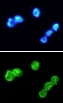
ARG56952 anti-IL33 antibody [4E9] ICC/IF image
Immunoflorescense: Jurkat cell line stained with ARG56952 anti-IL33 antibody [4E9] at 1:100 (Green).
DAPI (Blue) for nucleus staining.
-
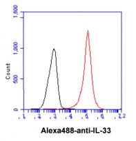
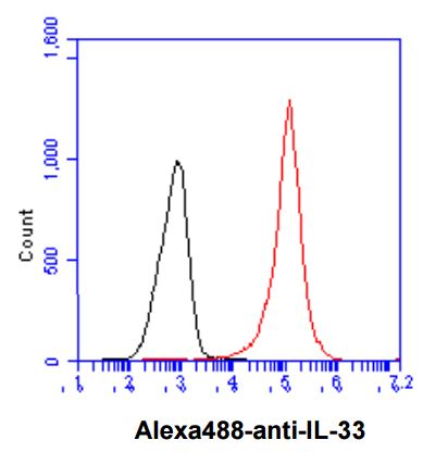
ARG56952 anti-IL33 antibody [4E9] FACS image
Flow Cytometry: Jurkat cell line stained with ARG56952 anti-IL33 antibody [4E9] at 2-5 µg for 1x10^6 cells (red line). Secondary antibody: Goat anti-Mouse IgG Alexa fluor 488 conjugate. Isotype control antibody was Mouse IgG (black line).
-
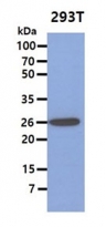
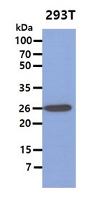
ARG56952 anti-IL33 antibody [4E9] WB image
Western blot: 40 µg of 293T cell lysate stained with ARG56952 anti-IL33 antibody [4E9] at 1:1000.
-
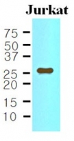
ARG56952 anti-IL33 antibody [4E9] WB image
Western blot: 35 µg of Jurkat cell lysate stained with ARG56952 anti-IL33 antibody [4E9] at 1:500.