ARG56599
anti-IL2 antibody [1H10-1-1]
anti-IL2 antibody [1H10-1-1] for ELISA,IHC-Formalin-fixed paraffin-embedded sections,Western blot and Human
Overview
| Product Description | Mouse Monoclonal antibody [1H10-1-1] recognizes IL2 |
|---|---|
| Tested Reactivity | Hu |
| Tested Application | ELISA, IHC-P, WB |
| Host | Mouse |
| Clonality | Monoclonal |
| Clone | 1H10-1-1 |
| Isotype | IgG1, kappa |
| Target Name | IL2 |
| Antigen Species | Human |
| Immunogen | E.coli derived Recombinant Human IL-2. (MAPTSSSTKK TQLQLEHLLL DLQMILNGIN NYKNPKLTRM LTFKFYMPKK ATELKHLQCL EEELKPLEEV LNLAQSKNFH LRPRDLISNI NVIVLELKGS ETTFMCEYAD ETATIVEFLN RWITFAQSII STLT) |
| Conjugation | Un-conjugated |
| Alternate Names | TCGF; IL-2; lymphokine; Interleukin-2; Aldesleukin; T-cell growth factor |
Application Instructions
| Application Suggestion |
|
||||||||
|---|---|---|---|---|---|---|---|---|---|
| Application Note | * The dilutions indicate recommended starting dilutions and the optimal dilutions or concentrations should be determined by the scientist. |
Properties
| Form | Liquid |
|---|---|
| Purification | Purification with Protein A. |
| Buffer | PBS (pH 7.2) |
| Concentration | 1 mg/ml |
| Storage Instruction | For continuous use, store undiluted antibody at 2-8°C for up to a week. For long-term storage, aliquot and store at -20°C or below. Storage in frost free freezers is not recommended. Avoid repeated freeze/thaw cycles. Suggest spin the vial prior to opening. The antibody solution should be gently mixed before use. |
| Note | For laboratory research only, not for drug, diagnostic or other use. |
Bioinformation
| Database Links | |
|---|---|
| Gene Symbol | IL2 |
| Gene Full Name | interleukin 2 |
| Background | The protein encoded by this gene is a secreted cytokine that is important for the proliferation of T and B lymphocytes. The receptor of this cytokine is a heterotrimeric protein complex whose gamma chain is also shared by interleukin 4 (IL4) and interleukin 7 (IL7). The expression of this gene in mature thymocytes is monoallelic, which represents an unusual regulatory mode for controlling the precise expression of a single gene. The targeted disruption of a similar gene in mice leads to ulcerative colitis-like disease, which suggests an essential role of this gene in the immune response to antigenic stimuli. [provided by RefSeq, Jul 2008] |
| Function | Produced by T-cells in response to antigenic or mitogenic stimulation, this protein is required for T-cell proliferation and other activities crucial to regulation of the immune response. Can stimulate B-cells, monocytes, lymphokine-activated killer cells, natural killer cells, and glioma cells. [UniProt] |
| Calculated MW | 18 kDa |
Images (4) Click the Picture to Zoom In
-

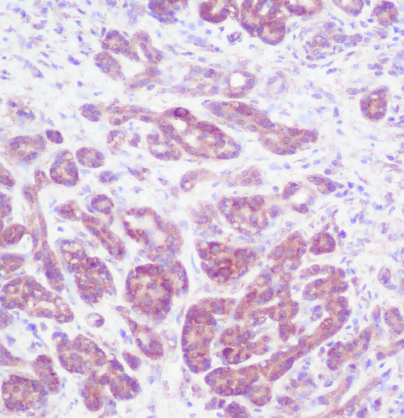
ARG56599 anti-IL2 antibody [1H10-1-1] IHC-P image
Immunohistochemistry: Formalin-fixed and paraffin-embedded sections of Human breast invasive ductal carcinoma. The recommended ARG56599 anti-IL2 antibody [1H10-1-1] concentration is 2.5 µg/ml-5.0 µg/ml with an overnight incubation at 4°C. An HRP-labeled polymer detection system was used with a DAB chromogen. Antigen Retrieval: Boil tissue section in Sodium Citrate buffer (pH 6.0) followed by cooling at RT for 20 min.
-
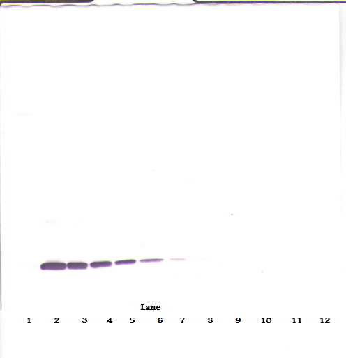
ARG56599 anti-IL2 antibody [1H10-1-1] WB image
Western blot: 250 - 0.24 ng (left to right) of Human IL-2 stained with ARG56599 anti-IL2 antibody [1H10-1-1], under reducing conditions.
-
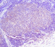
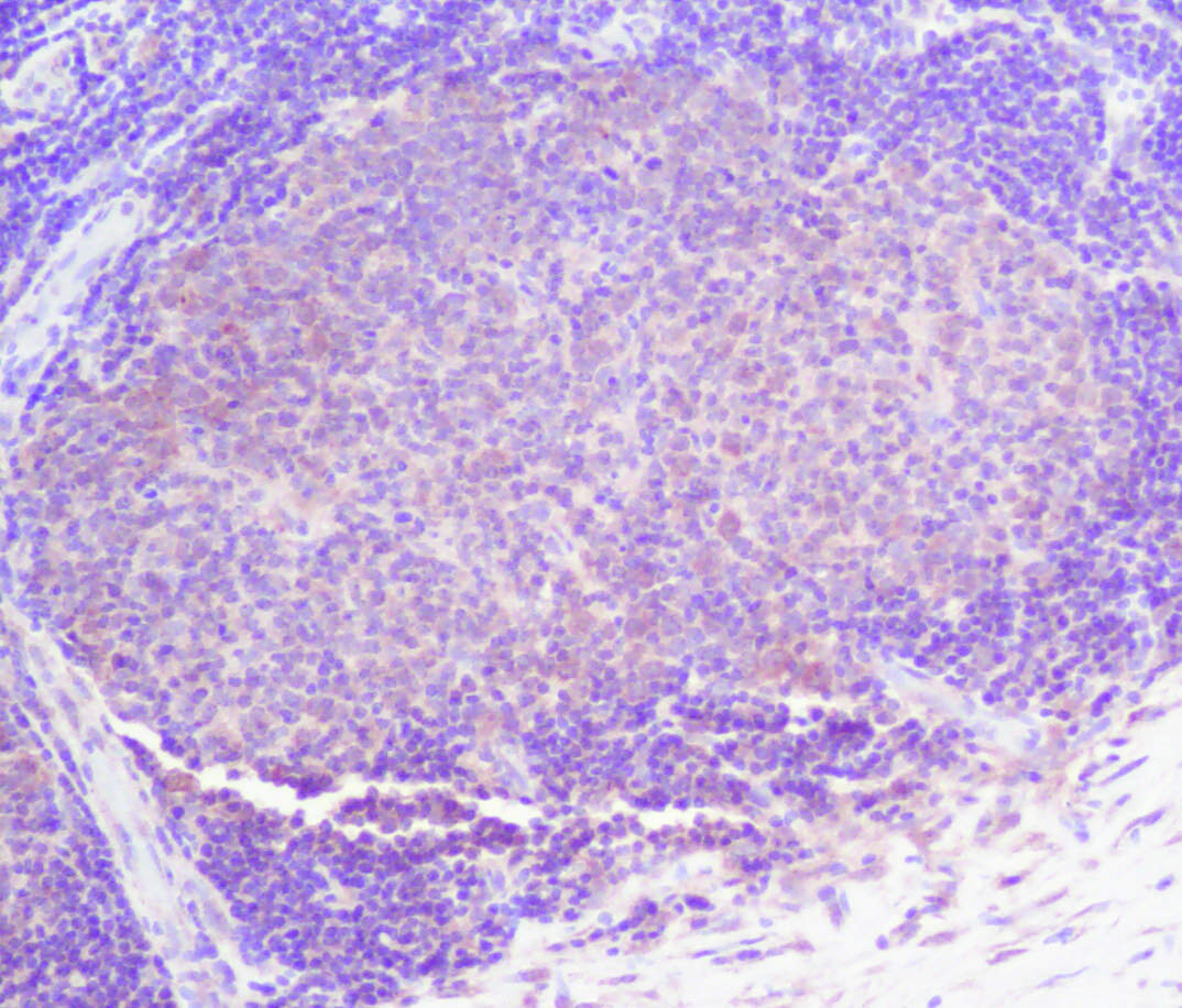
ARG56599 anti-IL2 antibody [1H10-1-1] IHC-P image
Immunohistochemistry: Formalin-fixed and paraffin-embedded sections of Human breast invasive ductal carcinoma. The recommended ARG56599 anti-IL2 antibody [1H10-1-1] concentration is 2.5 µg/ml-5.0 µg/ml with an overnight incubation at 4°C. An HRP-labeled polymer detection system was used with a DAB chromogen. Antigen Retrieval: Boil tissue section in Sodium Citrate buffer (pH 6.0) followed by cooling at RT for 20 min.
-
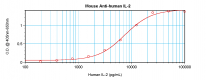
ARG56599 anti-IL2 antibody [1H10-1-1] standard curve image
Sandwich ELISA: ARG56599 anti-IL2 antibody [1H10-1-1] as a capture antibody at 4.0 - 8.0 µg/ml combined with anti-IL2 antibody (Biotin) as a detection antibody at ~ 1.0 - 2.0 µg/ml. Results of a typical standard run with optical density.