ARG66026
anti-IL17D antibody (Biotin)
anti-IL17D antibody (Biotin) for ELISA,Western blot and Human
Overview
| Product Description | Biotin-conjugated Rabbit Polyclonal antibody recognizes IL17D |
|---|---|
| Tested Reactivity | Hu |
| Tested Application | ELISA, WB |
| Host | Rabbit |
| Clonality | Polyclonal |
| Isotype | IgG |
| Target Name | IL17D |
| Antigen Species | Human |
| Immunogen | E. coli derived recombinant Human IL17D. (APRAGRRPAR PRGCADRPEE LLEQLYGRLA AGVLSAFHHT LQLGPREQAR NASCPAGGRP ADRRFRPPTN LRSVSPWAYR ISYDPARYPR YLPEAYCLCR GCLTGLFGEE DVRFRSAPVY MPTVVLRRTP ACAGGRSVYT EAYVTIPVGC TCVPEPEKDA DSINSSIDKQ GAKLLLGPND APAGP) |
| Conjugation | Biotin |
| Alternate Names | IL17D; Interleukin 17D; IL-17D; IL-27; Interleukin-17D; FLJ30846; IL27; Interleukin 27; Interleukin-27 |
Application Instructions
| Application Suggestion |
|
||||||
|---|---|---|---|---|---|---|---|
| Application Note | * The dilutions indicate recommended starting dilutions and the optimal dilutions or concentrations should be determined by the scientist. |
Properties
| Form | Liquid |
|---|---|
| Purification | Purified by affinity chromatography. |
| Buffer | PBS (pH 7.2) |
| Concentration | 1 mg/ml |
| Storage Instruction | Aliquot and store in the dark at 2-8°C. Keep protected from prolonged exposure to light. Avoid repeated freeze/thaw cycles. Suggest spin the vial prior to opening. The antibody solution should be gently mixed before use. |
| Note | For laboratory research only, not for drug, diagnostic or other use. |
Bioinformation
| Database Links | |
|---|---|
| Gene Symbol | IL17D |
| Gene Full Name | interleukin 17D |
| Background | The protein encoded by this gene is a cytokine that shares the sequence similarity with IL17. The treatment of endothelial cells with this cytokine has been shown to stimulate the production of other cytokines including IL6, IL8 and CSF2/ GM-CSF. The increased expression of IL8 induced by this cytokine was found to be NF-kappa B-dependent. [provided by RefSeq, Jul 2008] |
| Function | Induces expression of IL-6, IL-8, and GM-CSF from endothelial cells. [UniProt] |
| Calculated MW | 22 kDa |
Images (4) Click the Picture to Zoom In
-
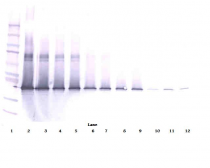
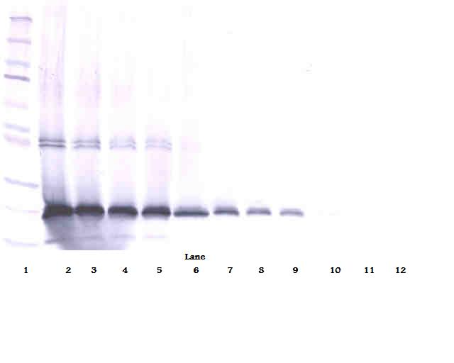
ARG66026 anti-IL17D antibody (Biotin) WB image
Western blot: 250 - 0.24 ng of Human IL-17D stained with ARG66026 anti-IL17D antibody (Biotin), under non-reducing conditions.
-
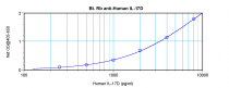
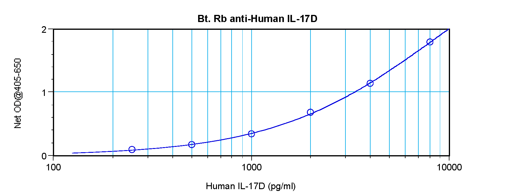
ARG66026 anti-IL17D antibody (Biotin) standard curve image
Direct ELISA: ARG66026 anti-IL17D antibody (Biotin) at ~ 1.0 µg/ml results of a typical standard run with optical density reading at 405 - 650 nm.
-
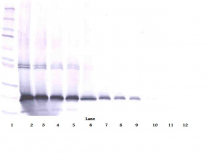
ARG66026 anti-IL17D antibody (Biotin) WB image
Western blot: 250 - 0.24 ng of Human IL-17D stained with ARG66026 anti-IL17D antibody (Biotin), under reducing conditions.
-
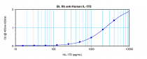
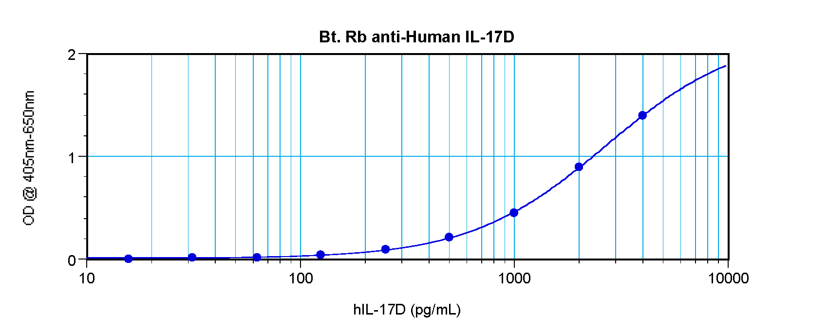
ARG66026 anti-IL17D antibody (Biotin) standard curve image
Sandwich ELISA: ARG66026 anti-IL17D antibody (Biotin) as a detection antibody at 0.25 - 1.0 µg/ml combined with ARG66025 anti-IL17D antibody as a capture antibody. Results of a typical standard run with optical density reading at 405 - 650 nm.