ARG42282
anti-IKZF1 / Ikaros antibody [4E9] (PE)
anti-IKZF1 / Ikaros antibody [4E9] (PE) for Flow cytometry and Human,Mouse
Overview
| Product Description | PE-conjugated Mouse Monoclonal antibody [4E9] recognizes IKZF1 / Ikaros |
|---|---|
| Tested Reactivity | Hu, Ms |
| Tested Application | FACS |
| Specificity | The mouse monoclonal antibody 4E9 recognizes Ikaros, a transcription factor (intracellular antigen) expressed broadly in hematopoietic progenitors and serving as a key regulator of lymphopoiesis. |
| Host | Mouse |
| Clonality | Monoclonal |
| Clone | 4E9 |
| Isotype | IgG1 |
| Target Name | IKZF1 / Ikaros |
| Antigen Species | Human |
| Immunogen | Recombinant Human Ikaros (C-terminal part). |
| Conjugation | PE |
| Alternate Names | IK1; Hs.54452; LYF1; PPP1R92; LyF-1; Lymphoid transcription factor LyF-1; IKAROS; Ikaros family zinc finger protein 1; DNA-binding protein Ikaros; PRO0758; ZNFN1A1 |
Application Instructions
| Application Suggestion |
|
||||
|---|---|---|---|---|---|
| Application Note | * The dilutions indicate recommended starting dilutions and the optimal dilutions or concentrations should be determined by the scientist. |
Properties
| Form | Liquid |
|---|---|
| Purification | Purified |
| Buffer | PBS and 15 mM Sodium azide. |
| Preservative | 15 mM Sodium azide |
| Storage Instruction | Aliquot and store in the dark at 2-8°C. Keep protected from prolonged exposure to light. Avoid repeated freeze/thaw cycles. Suggest spin the vial prior to opening. The antibody solution should be gently mixed before use. |
| Note | For laboratory research only, not for drug, diagnostic or other use. |
Bioinformation
| Database Links | |
|---|---|
| Gene Symbol | IKZF1 |
| Gene Full Name | IKAROS family zinc finger 1 (Ikaros) |
| Background | This gene encodes a transcription factor that belongs to the family of zinc-finger DNA-binding proteins associated with chromatin remodeling. The expression of this protein is restricted to the fetal and adult hemo-lymphopoietic system, and it functions as a regulator of lymphocyte differentiation. Several alternatively spliced transcript variants encoding different isoforms have been described for this gene. Most isoforms share a common C-terminal domain, which contains two zinc finger motifs that are required for hetero- or homo-dimerization, and for interactions with other proteins. The isoforms, however, differ in the number of N-terminal zinc finger motifs that bind DNA and in nuclear localization signal presence, resulting in members with and without DNA-binding properties. Only a few isoforms contain the requisite three or more N-terminal zinc motifs that confer high affinity binding to a specific core DNA sequence element in the promoters of target genes. The non-DNA-binding isoforms are largely found in the cytoplasm, and are thought to function as dominant-negative factors. Overexpression of some dominant-negative isoforms have been associated with B-cell malignancies, such as acute lymphoblastic leukemia (ALL). [provided by RefSeq, May 2014] |
| Function | Transcription regulator of hematopoietic cell differentiation (PubMed:17934067). Binds gamma-satellite DNA (PubMed:17135265, PubMed:19141594). Plays a role in the development of lymphocytes, B- and T-cells. Binds and activates the enhancer (delta-A element) of the CD3-delta gene. Repressor of the TDT (fikzfterminal deoxynucleotidyltransferase) gene during thymocyte differentiation. Regulates transcription through association with both HDAC-dependent and HDAC-independent complexes. Targets the 2 chromatin-remodeling complexes, NuRD and BAF (SWI/SNF), in a single complex (PYR complex), to the beta-globin locus in adult erythrocytes. Increases normal apoptosis in adult erythroid cells. Confers early temporal competence to retinal progenitor cells (RPCs) (By similarity). Function is isoform-specific and is modulated by dominant-negative inactive isoforms (PubMed:17135265, PubMed:17934067). [UniProt] |
| Cellular Localization | Nucleus. Note=In resting lymphocytes, distributed diffusely throughout the nucleus. Localizes to pericentromeric heterochromatin in proliferating cells. This localization requires DNA binding which is regulated by phosphorylation / dephosphorylation events. Isoform Ik2: Nucleus. Isoform Ik6: Cytoplasm. [UniProt] |
| Calculated MW | 58 kDa |
| PTM | Phosphorylation controls cell-cycle progression from late G(1) stage to S stage. Hyperphosphorylated during G2/M phase. Dephosphorylated state during late G(1) phase. Phosphorylation on Thr-140 is required for DNA and pericentromeric location during mitosis. CK2 is the main kinase, in vitro. GSK3 and CDK may also contribute to phosphorylation of the C-terminal serine and threonine residues. Phosphorylation on these C-terminal residues reduces the DNA-binding ability. Phosphorylation/dephosphorylation events on Ser-13 and Ser-295 regulate TDT expression during thymocyte differentiation. Dephosphorylation by protein phosphatase 1 regulates stability and pericentromeric heterochromatin location. Phosphorylated in both lymphoid and non-lymphoid tissues (By similarity). Phosphorylation at Ser-361 and Ser-364 downstream of SYK induces nuclear translocation. Sumoylated. Simulataneous sumoylation on the 2 sites results in a loss of both HDAC-dependent and HDAC-independent repression. Has no effect on pericentromeric heterochromatin location. Desumoylated by SENP1 (By similarity). Polyubiquitinated. [UniProt] |
Images (1) Click the Picture to Zoom In
-
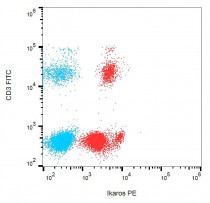
ARG42282 anti-IKZF1 / Ikaros antibody [4E9] (PE) FACS image
Flow Cytometry: Human peripheral blood leukocytes stained with ARG42282 anti-IKZF1 / Ikaros antibody [4E9] (PE) and anti-CD3 antibody (FITC).


