ARG40919
anti-IGFBP2 antibody
anti-IGFBP2 antibody for Flow cytometry,ICC/IF,IHC-Formalin-fixed paraffin-embedded sections,Western blot and Mouse,Rat
Overview
| Product Description | Rabbit Polyclonal antibody recognizes IGFBP2 |
|---|---|
| Tested Reactivity | Ms, Rat |
| Tested Application | FACS, ICC/IF, IHC-P, WB |
| Host | Rabbit |
| Clonality | Polyclonal |
| Isotype | IgG |
| Target Name | IGFBP2 |
| Antigen Species | Rat |
| Immunogen | Recombinant protein corresponding to E35-Q304 of Rat IGFBP2. |
| Conjugation | Un-conjugated |
| Alternate Names | IBP-2; IBP2; Insulin-like growth factor-binding protein 2; IGFBP-2; IGF-BP53; IGF-binding protein 2 |
Application Instructions
| Application Suggestion |
|
||||||||||
|---|---|---|---|---|---|---|---|---|---|---|---|
| Application Note | IHC-P: Antigen Retrieval: Heat mediation was performed in Citrate buffer (pH 6.0) for 20 min. * The dilutions indicate recommended starting dilutions and the optimal dilutions or concentrations should be determined by the scientist. |
Properties
| Form | Liquid |
|---|---|
| Purification | Affinity purification with immunogen. |
| Buffer | 0.2% Na2HPO4, 0.9% NaCl, 0.05% Sodium azide and 4% Trehalose. |
| Preservative | 0.05% Sodium azide |
| Stabilizer | 4% Trehalose |
| Concentration | 0.5 mg/ml |
| Storage Instruction | For continuous use, store undiluted antibody at 2-8°C for up to a week. For long-term storage, aliquot and store at -20°C or below. Storage in frost free freezers is not recommended. Avoid repeated freeze/thaw cycles. Suggest spin the vial prior to opening. The antibody solution should be gently mixed before use. |
| Note | For laboratory research only, not for drug, diagnostic or other use. |
Bioinformation
| Database Links |
Swiss-port # P12843 Rat Insulin-like growth factor-binding protein 2 Swiss-port # P47877 Mouse Insulin-like growth factor-binding protein 2 |
|---|---|
| Gene Symbol | IGFBP2 |
| Gene Full Name | insulin-like growth factor binding protein 2, 36kDa |
| Background | The protein encoded by this gene is one of six similar proteins that bind insulin-like growth factors I and II (IGF-I and IGF-II). The encoded protein can be secreted into the bloodstream, where it binds IGF-I and IGF-II with high affinity, or it can remain intracellular, interacting with many different ligands. High expression levels of this protein promote the growth of several types of tumors and may be predictive of the chances of recovery of the patient. Several transcript variants, one encoding a secreted isoform and the others encoding nonsecreted isoforms, have been found for this gene. [provided by RefSeq, Sep 2015] |
| Function | Inhibits IGF-mediated growth and developmental rates. IGF-binding proteins prolong the half-life of the IGFs and have been shown to either inhibit or stimulate the growth promoting effects of the IGFs on cell culture. They alter the interaction of IGFs with their cell surface receptors. [UniProt] |
| Cellular Localization | Secreted. [UniProt] |
| Calculated MW | 35 kDa |
| PTM | O-glycosylated. [UniProt] |
Images (8) Click the Picture to Zoom In
-

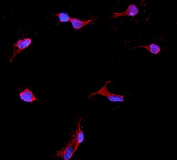
ARG40919 anti-IGFBP2 antibody ICC/IF image
Immunofluorescence: NRK cells were blocked with 10% goat serum and then stained with ARG40919 anti-IGFBP2 antibody (red) at 2 µg/ml dilution, overnight at 4°C. DAPI (blue) for nuclear staining.
-
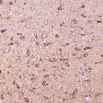
ARG40919 anti-IGFBP2 antibody IHC-P image
Immunohistochemistry: Paraffin-embedded Mouse brain tissue. Antigen Retrieval: Heat mediation was performed in Citrate buffer (pH 6.0, epitope retrieval solution) for 20 min. The tissue section was blocked with 10% goat serum. The tissue section was then stained with ARG40919 anti-IGFBP2 antibody at 2 ug/ml, overnight at 4°C.
-
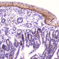
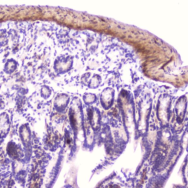
ARG40919 anti-IGFBP2 antibody IHC-P image
Immunohistochemistry: Paraffin-embedded Mouse small intestine tissue. Antigen Retrieval: Heat mediation was performed in Citrate buffer (pH 6.0) for 20 min. The tissue section was blocked with 10% goat serum. The tissue section was then stained with ARG40919 anti-IGFBP2 antibody at 2 µg/ml dilution, overnight at 4°C.
-
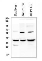
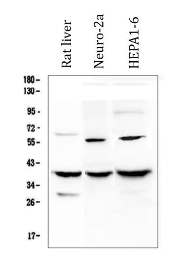
ARG40919 anti-IGFBP2 antibody WB image
Western blot: 50 ug of samples under reducing conditions. Rat liver, Neuro-2a and HEPA1-6 whole cell lysates stained with ARG40919 anti-IGFBP2 antibody at 0.5 ug/mL, overnight at 4°C.
-
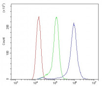
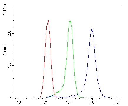
ARG40919 anti-IGFBP2 antibody FACS image
Flow Cytometry: RH35 cells were blocked with 10% normal goat serum and then stained with ARG40919 anti-IGFBP2 antibody (blue) at 1 µg/10^6 cells for 30 min at 20°C, followed by incubation with DyLight®488 labelled secondary antibody. Isotype control antibody (green) was rabbit IgG (1 µg/10^6 cells) used under the same conditions. Unlabelled sample (red) was also used as a control.
-
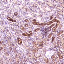
ARG40919 anti-IGFBP2 antibody IHC-P image
Immunohistochemistry: Paraffin-embedded Mouse kidney tissue . Antigen Retrieval: Heat mediation was performed in Citrate buffer (pH 6.0) for 20 min. The tissue section was blocked with 10% goat serum. The tissue section was then stained with ARG40919 anti-IGFBP2 antibody at 2 µg/ml dilution, overnight at 4°C.
-
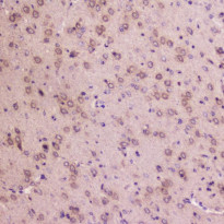
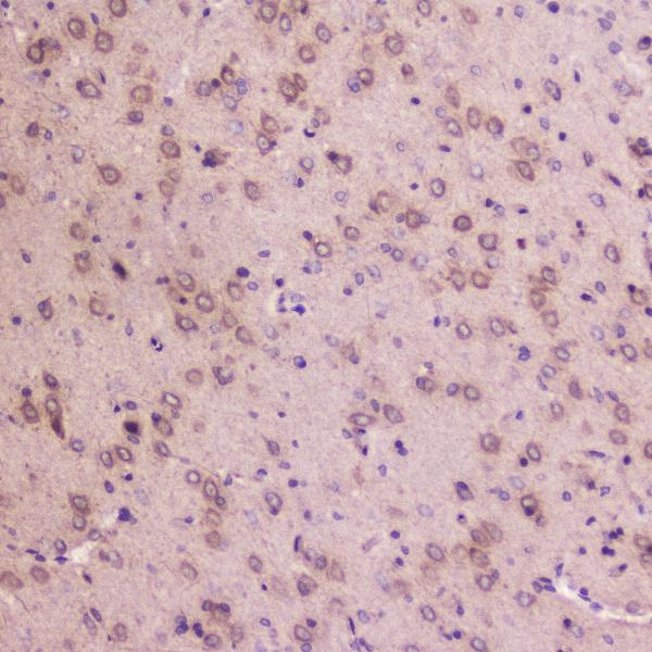
ARG40919 anti-IGFBP2 antibody IHC-P image
Immunohistochemistry: Paraffin-embedded Rat brain tissue . Antigen Retrieval: Heat mediation was performed in Citrate buffer (pH 6.0) for 20 min. The tissue section was blocked with 10% goat serum. The tissue section was then stained with ARG40919 anti-IGFBP2 antibody at 2 µg/ml dilution, overnight at 4°C.
-
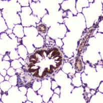
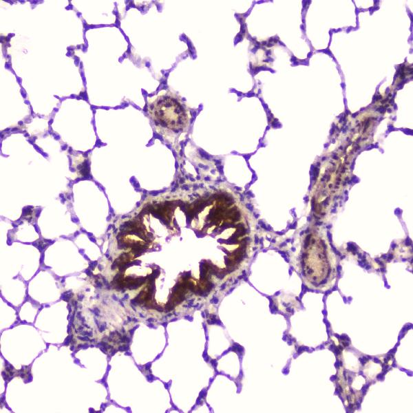
ARG40919 anti-IGFBP2 antibody IHC-P image
Immunohistochemistry: Paraffin-embedded Rat lung tissue . Antigen Retrieval: Heat mediation was performed in Citrate buffer (pH 6.0) for 20 min. The tissue section was blocked with 10% goat serum. The tissue section was then stained with ARG40919 anti-IGFBP2 antibody at 2 µg/ml dilution, overnight at 4°C.