ARG40686
anti-IGFBP1 antibody
anti-IGFBP1 antibody for IHC-Formalin-fixed paraffin-embedded sections,Western blot and Human
Overview
| Product Description | Rabbit Polyclonal antibody recognizes IGFBP1 |
|---|---|
| Tested Reactivity | Hu |
| Tested Application | IHC-P, WB |
| Host | Rabbit |
| Clonality | Polyclonal |
| Isotype | IgG |
| Target Name | IGFBP1 |
| Antigen Species | Human |
| Immunogen | Recombinant protein corresponding to A69-N259 of Human IGFBP1. |
| Conjugation | Un-conjugated |
| Alternate Names | IBP-1; IBP1; PP12; IGF-BP25; Insulin-like growth factor-binding protein 1; hIGFBP-1; IGFBP-1; Placental protein 12; AFBP; IGF-binding protein 1 |
Application Instructions
| Application Suggestion |
|
||||||
|---|---|---|---|---|---|---|---|
| Application Note | IHC-P: Antigen Retrieval: Heat mediation was performed in Citrate buffer (pH 6.0, epitope retrieval solution) for 20 min. * The dilutions indicate recommended starting dilutions and the optimal dilutions or concentrations should be determined by the scientist. |
Properties
| Form | Liquid |
|---|---|
| Purification | Affinity purification with immunogen. |
| Buffer | 0.2% Na2HPO4, 0.9% NaCl, 0.05% Sodium azide and 5% BSA. |
| Preservative | 0.05% Sodium azide |
| Stabilizer | 5% BSA |
| Concentration | 0.5 mg/ml |
| Storage Instruction | For continuous use, store undiluted antibody at 2-8°C for up to a week. For long-term storage, aliquot and store at -20°C or below. Storage in frost free freezers is not recommended. Avoid repeated freeze/thaw cycles. Suggest spin the vial prior to opening. The antibody solution should be gently mixed before use. |
| Note | For laboratory research only, not for drug, diagnostic or other use. |
Bioinformation
| Database Links |
Swiss-port # P08833 Human Insulin-like growth factor-binding protein 1 |
|---|---|
| Gene Symbol | IGFBP1 |
| Gene Full Name | insulin-like growth factor binding protein 1 |
| Background | This gene is a member of the insulin-like growth factor binding protein (IGFBP) family and encodes a protein with an IGFBP domain and a thyroglobulin type-I domain. The protein binds both insulin-like growth factors (IGFs) I and II and circulates in the plasma. Binding of this protein prolongs the half-life of the IGFs and alters their interaction with cell surface receptors. [provided by RefSeq, Jul 2008] |
| Function | IGF-binding proteins prolong the half-life of the IGFs and have been shown to either inhibit or stimulate the growth promoting effects of the IGFs on cell culture. They alter the interaction of IGFs with their cell surface receptors. Promotes cell migration. [UniProt] |
| Cellular Localization | Secreted. [UniProt] |
| Calculated MW | 28 kDa |
| PTM | Phosphorylated; probably by casein kinase II. Phosphorylation alters the affinity of the protein for IGFs. In amniotic fluid, the unmodified protein is the most abundant form, while mono-, bi-, tri- and tetraphosphorylated forms are present in decreasing amounts. The phosphorylation state may influence the propensity to proteolysis. [UniProt] |
Images (2) Click the Picture to Zoom In
-

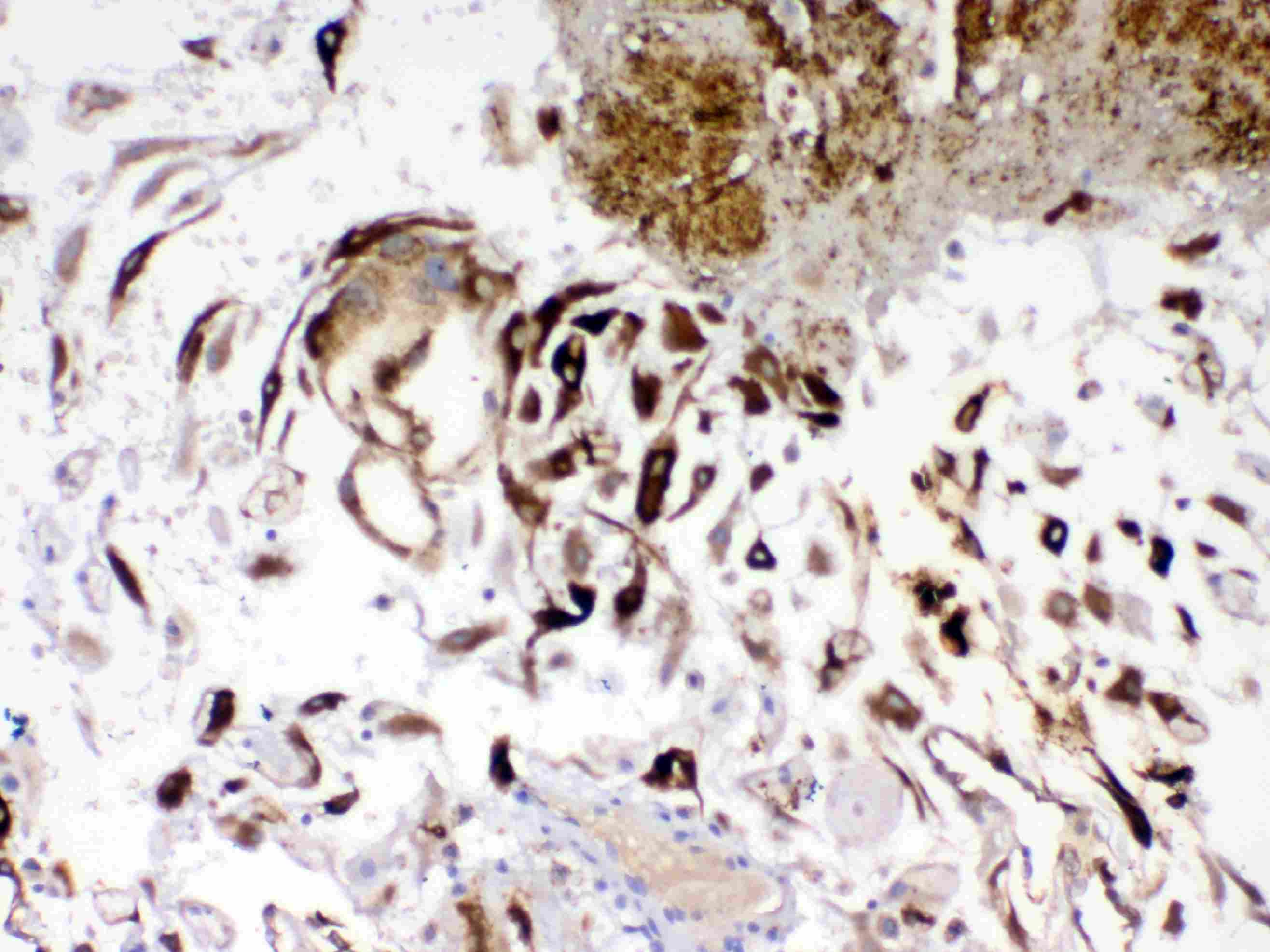
ARG40686 anti-IGFBP1 antibody IHC-P image
Immunohistochemistry: Paraffin-embedded Human placenta tissue. Antigen Retrieval: Heat mediation was performed in Citrate buffer (pH 6.0, epitope retrieval solution) for 20 min. The tissue section was blocked with 10% goat serum. The tissue section was then stained with ARG40686 anti-IGFBP1 antibody at 1 µg/ml, overnight at 4°C.
-
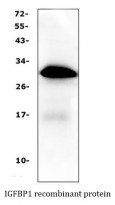
ARG40686 anti-IGFBP1 antibody WB image
Western blot: IGFBP1 recombinant protein stained with ARG40686 anti-IGFBP1 antibody.