ARG42968
anti-IFNAR2 antibody
anti-IFNAR2 antibody for Flow cytometry,IHC-Formalin-fixed paraffin-embedded sections,Western blot and Human,Mouse,Rat
Overview
| Product Description | Rabbit Polyclonal antibody recognizes IFNAR2 |
|---|---|
| Tested Reactivity | Hu, Ms, Rat |
| Tested Application | FACS, IHC-P, WB |
| Host | Rabbit |
| Clonality | Polyclonal |
| Isotype | IgG |
| Target Name | IFNAR2 |
| Antigen Species | Human |
| Immunogen | Recombinant protein corresponding to D33-Q466 of Human IFNAR2. |
| Conjugation | Un-conjugated |
| Alternate Names | IFNABR; IFNARB; IFN-R; IFN-alpha/beta receptor 2; IFN-R-2; Type I interferon receptor 2; Interferon alpha binding protein; IFN-alpha binding protein; IFN-alpha-REC; Interferon alpha/beta receptor 2 |
Application Instructions
| Application Suggestion |
|
||||||||
|---|---|---|---|---|---|---|---|---|---|
| Application Note | IHC-P: Antigen Retrieval: Heat mediation was performed in Citrate buffer (pH 6.0) for 20 min. * The dilutions indicate recommended starting dilutions and the optimal dilutions or concentrations should be determined by the scientist. |
||||||||
| Observed Size | ~ 65 kDa |
Properties
| Form | Liquid |
|---|---|
| Purification | Affinity purification with immunogen. |
| Buffer | 0.2% Na2HPO4, 0.9% NaCl, 0.05% Sodium azide and 4% Trehalose. |
| Preservative | 0.05% Sodium azide |
| Stabilizer | 4% Trehalose |
| Concentration | 0.5 mg/ml |
| Storage Instruction | For continuous use, store undiluted antibody at 2-8°C for up to a week. For long-term storage, aliquot and store at -20°C or below. Storage in frost free freezers is not recommended. Avoid repeated freeze/thaw cycles. Suggest spin the vial prior to opening. The antibody solution should be gently mixed before use. |
| Note | For laboratory research only, not for drug, diagnostic or other use. |
Bioinformation
| Database Links | |
|---|---|
| Gene Symbol | IFNAR2 |
| Gene Full Name | interferon (alpha, beta and omega) receptor 2 |
| Background | The protein encoded by this gene is a type I membrane protein that forms one of the two chains of a receptor for interferons alpha and beta. Binding and activation of the receptor stimulates Janus protein kinases, which in turn phosphorylate several proteins, including STAT1 and STAT2. Multiple transcript variants encoding at least two different isoforms have been found for this gene. [provided by RefSeq, Jul 2008] |
| Function | Associates with IFNAR1 to form the type I interferon receptor. Receptor for interferons alpha and beta. Involved in IFN-mediated STAT1, STAT2 and STAT3 activation (PubMed:26424569). Isoform 1 and isoform 2 are directly involved in signal transduction due to their association with the TYR kinase, JAK1 (PubMed:8181059, PubMed:7665574, PubMed:7759950). Isoform 3 is a potent inhibitor of type I IFN receptor activity (PubMed:7759950). [UniProt] |
| Cellular Localization | Isoform 1: Cell membrane; Single-pass type I membrane protein. Isoform 2: Cell membrane; Single-pass type I membrane protein. Isoform 3: Secreted. [UniProt] |
| Calculated MW | 58 kDa |
| PTM | Phosphorylated on tyrosine residues upon interferon binding. Phosphorylation at Tyr-337 or Tyr-512 are sufficient to mediate interferon dependent activation of STAT1, STAT2 and STAT3 leading to antiproliferative effects on many different cell types. Glycosylated. [UniProt] |
Images (6) Click the Picture to Zoom In
-

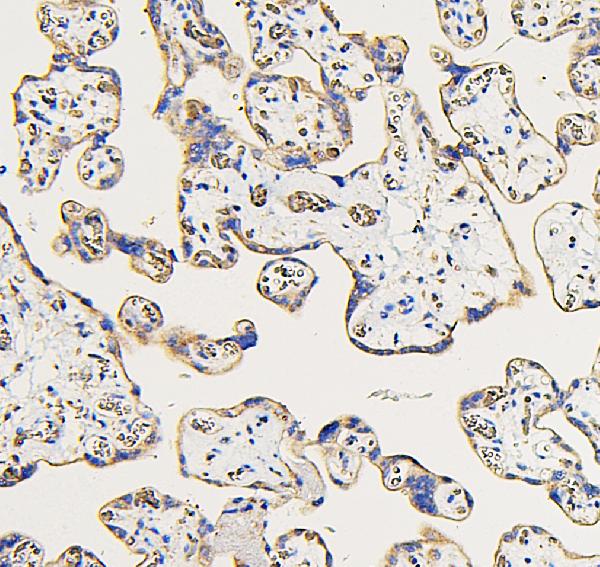
ARG42968 anti-IFNAR2 antibody IHC-P image
Immunohistochemistry: Paraffin-embedded Human placenta tissue. Antigen Retrieval: Heat mediation was performed in Citrate buffer (pH 6.0) for 20 min. The tissue section was blocked with 10% goat serum. The tissue section was then stained with ARG42968 anti-IFNAR2 antibody at 1 µg/ml dilution, overnight at 4°C.
-
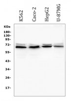
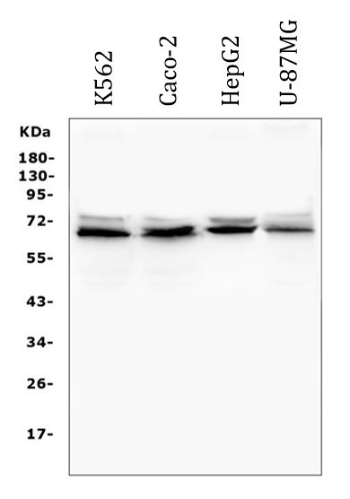
ARG42968 anti-IFNAR2 antibody WB image
Western blot: 50 µg of sample under reducing conditions. K562, Caco-2, HepG2 and U-87MG whole cell lysates stained with ARG42968 anti-IFNAR2 antibody at 0.5 µg/ml dilution, overnight at 4°C.
-
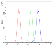
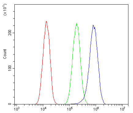
ARG42968 anti-IFNAR2 antibody FACS image
Flow Cytometry: A549 cells were blocked with 10% normal goat serum and then stained with ARG42968 anti-IFNAR2 antibody (blue) at 1 µg/10^6 cells for 30 min at 20°C, followed by incubation with DyLight®488 labelled secondary antibody. Isotype control antibody (green) was Rabbit IgG (1 µg/10^6 cells) used under the same conditions. Unlabelled sample (red) was also used as a control.
-

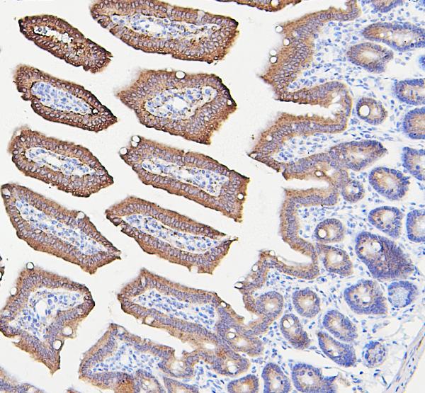
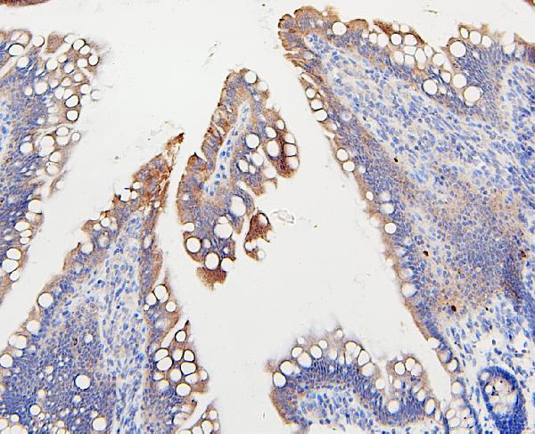
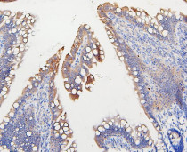
ARG42968 anti-IFNAR2 antibody IHC-P image
Immunohistochemistry: Paraffin-embedded Mouse intestine tissue. Antigen Retrieval: Heat mediation was performed in Citrate buffer (pH 6.0) for 20 min. The tissue section was blocked with 10% goat serum. The tissue section was then stained with ARG42968 anti-IFNAR2 antibody at 1 µg/ml dilution, overnight at 4°C.
-

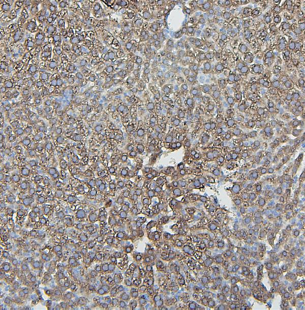
ARG42968 anti-IFNAR2 antibody IHC-P image
Immunohistochemistry: Paraffin-embedded Mouse liver tissue. Antigen Retrieval: Heat mediation was performed in Citrate buffer (pH 6.0) for 20 min. The tissue section was blocked with 10% goat serum. The tissue section was then stained with ARG42968 anti-IFNAR2 antibody at 1 µg/ml dilution, overnight at 4°C.
-
ARG42968 anti-IFNAR2 antibody IHC-P image
Immunohistochemistry: Paraffin-embedded Rat intestine tissue. Antigen Retrieval: Heat mediation was performed in Citrate buffer (pH 6.0) for 20 min. The tissue section was blocked with 10% goat serum. The tissue section was then stained with ARG42968 anti-IFNAR2 antibody at 1 µg/ml dilution, overnight at 4°C.