ARG44677
anti-IFIH1 / MDA5 antibody
anti-IFIH1 / MDA5 antibody for IHC-Formalin-fixed paraffin-embedded sections,Immunoprecipitation,Western blot and Human
Overview
| Product Description | Mouse Monoclonal antibody recognizes IFIH1 / MDA5 |
|---|---|
| Tested Reactivity | Hu |
| Tested Application | IHC-P, IP, WB |
| Host | Mouse |
| Clonality | Monoclonal |
| Isotype | IgG2a |
| Target Name | IFIH1 / MDA5 |
| Antigen Species | Human |
| Conjugation | Un-conjugated |
| Alternate Names | Interferon-induced helicase C domain-containing protein 1; Murabutide down-regulated protein; SGMRT1; EC 3.6.4.13; RIG-I-like receptor 2; MDA5; Clinically amyopathic dermatomyositis autoantigen 140 kDa; AGS7; MDA-5; CADM-140 autoantigen; Melanoma differentiation-associated protein 5; IDDM19; Interferon-induced with helicase C domain protein 1; RNA helicase-DEAD box protein 116; Helicard; RLR-2; Hlcd; Helicase with 2 CARD domains |
Application Instructions
| Application Suggestion |
|
||||||||
|---|---|---|---|---|---|---|---|---|---|
| Application Note | * The dilutions indicate recommended starting dilutions and the optimal dilutions or concentrations should be determined by the scientist. |
Properties
| Form | Liquid |
|---|---|
| Purification | Protein A purification |
| Buffer | PBS with 0.09% sodium azide |
| Storage Instruction | For continuous use, store undiluted antibody at 2-8°C for up to a week. For long-term storage, aliquot and store at -20°C or below. Storage in frost free freezers is not recommended. Avoid repeated freeze/thaw cycles. Suggest spin the vial prior to opening. The antibody solution should be gently mixed before use. |
| Note | For laboratory research only, not for drug, diagnostic or other use. |
Bioinformation
| Database Links |
Swiss-port # Q9BYX4 Human Interferon-induced helicase C domain-containing protein 1 |
|---|---|
| Gene Symbol | IFIH1 |
| Gene Full Name | interferon induced with helicase C domain 1 |
| Background | DEAD box proteins, characterized by the conserved motif Asp-Glu-Ala-Asp (DEAD), are putative RNA helicases. They are implicated in a number of cellular processes involving alteration of RNA secondary structure such as translation initiation, nuclear and mitochondrial splicing, and ribosome and spliceosome assembly. Based on their distribution patterns, some members of this family are believed to be involved in embryogenesis, spermatogenesis, and cellular growth and division. This gene encodes a DEAD box protein that is upregulated in response to treatment with beta-interferon and a protein kinase C-activating compound, mezerein. Irreversible reprogramming of melanomas can be achieved by treatment with both these agents; treatment with either agent alone only achieves reversible differentiation. Genetic variation in this gene is associated with diabetes mellitus insulin-dependent type 19. [provided by RefSeq, Jul 2012] |
| Function | Innate immune receptor which acts as a cytoplasmic sensor of viral nucleic acids and plays a major role in sensing viral infection and in the activation of a cascade of antiviral responses including the induction of type I interferons and proinflammatory cytokines. Its ligands include mRNA lacking 2'-O-methylation at their 5' cap and long-dsRNA (>1 kb in length). Upon ligand binding it associates with mitochondria antiviral signaling protein (MAVS/IPS1) which activates the IKK-related kinases: TBK1 and IKBKE which phosphorylate interferon regulatory factors: IRF3 and IRF7 which in turn activate transcription of antiviral immunological genes, including interferons (IFNs); IFN-alpha and IFN-beta. Responsible for detecting the Picornaviridae family members such as encephalomyocarditis virus (EMCV) and mengo encephalomyocarditis virus (ENMG). Can also detect other viruses such as dengue virus (DENV), west Nile virus (WNV), and reovirus. Also involved in antiviral signaling in response to viruses containing a dsDNA genome, such as vaccinia virus. Plays an important role in amplifying innate immune signaling through recognition of RNA metabolites that are produced during virus infection by ribonuclease L (RNase L). May play an important role in enhancing natural killer cell function and may be involved in growth inhibition and apoptosis in several tumor cell lines. [UniProt] |
| Cellular Localization | Cytoplasm. Nucleus. Note=May be found in the nucleus, during apoptosis. [UniProt] |
| Calculated MW | 117 kDa |
| PTM | N-glycosylation enhances cell surface expression and lengthens receptor half-life by preventing degradation in the ER. |
Images (3) Click the Picture to Zoom In
-
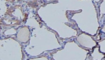
ARG44677 anti-IFIH1 / MDA5 antibody IHC-P image
Immunohistochemistry: Human Thyroid Gland stained with ARG44677 anti-IFIH1 / MDA5 antibody at 5 µg/mL dilution.
-
ARG44677 anti-IFIH1 / MDA5 antibody WB image
Western blot: THP1(WT) and THP1 (treated 15 µg LPS) stained with ARG44677 anti-IFIH1 / MDA5 antibody at 1 µg/mL dilution.
-
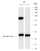
ARG44677 anti-IFIH1 / MDA5 antibody IP image
Immunoprecipitation: THP1 lysate immunoprecipitated with 2.5 µg of ARG44677 anti-IFIH1 / MDA5 antibody.


