ARG44438
anti-IDUA antibody
anti-IDUA antibody for IHC-Formalin-fixed paraffin-embedded sections,Western blot,Flow cytometry and Human,Mouse,Rat
Overview
| Product Description | Rabbit Polyclonal antibody recognizes IDUA |
|---|---|
| Tested Reactivity | Hu, Ms, Rat |
| Tested Application | FACS, IHC-P, WB |
| Host | Rabbit |
| Clonality | Polyclonal |
| Target Name | IDUA |
| Antigen Species | Human |
| Immunogen | Human IDUA recombinant protein |
| Conjugation | Un-conjugated |
| Alternate Names | IDUA; Alpha-L-Iduronidase; MPS1; MPSI; Mucopolysaccharidosis Type I |
Application Instructions
| Application Suggestion |
|
||||||||
|---|---|---|---|---|---|---|---|---|---|
| Application Note | The dilutions indicate recommended starting dilutions and the optimal dilutions or concentrations should be determined by the scientist. |
Properties
| Form | Liquid |
|---|---|
| Purification | Affinity purification with immunogen. |
| Buffer | 0.9% NaCl, 0.2% Na2HPO4, 0.05% Sodium azide and 4% Trehalose. |
| Preservative | 0.05% Sodium azide |
| Stabilizer | 4% Trehalose |
| Concentration | 0.5 mg/ml |
| Storage Instruction | For continuous use, store undiluted antibody at 2-8°C for up to a week. For long-term storage, aliquot and store at -20°C or below. Storage in frost free freezers is not recommended. Avoid repeated freeze/thaw cycles. Suggest spin the vial prior to opening. The antibody solution should be gently mixed before use. |
Bioinformation
| Database Links | |
|---|---|
| Gene Symbol | IDUA |
| Gene Full Name | Alpha-L-Iduronidase |
| Background | This gene encodes an enzyme that hydrolyzes the terminal alpha-L-iduronic acid residues of two glycosaminoglycans, dermatan sulfate and heparan sulfate. This hydrolysis is required for the lysosomal degradation of these glycosaminoglycans. Mutations in this gene that result in enzymatic deficiency lead to the autosomal recessive disease mucopolysaccharidosis type I (MPS I). |
| Function | Hydrolysis of unsulfated alpha-L-iduronosidic linkages in dermatan sulfate. |
| Cellular Localization | Lysosome |
| Calculated MW | 73 kDa |
| PTM | Disulfide bond, Glycoprotein |
Images (8) Click the Picture to Zoom In
-
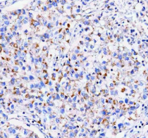
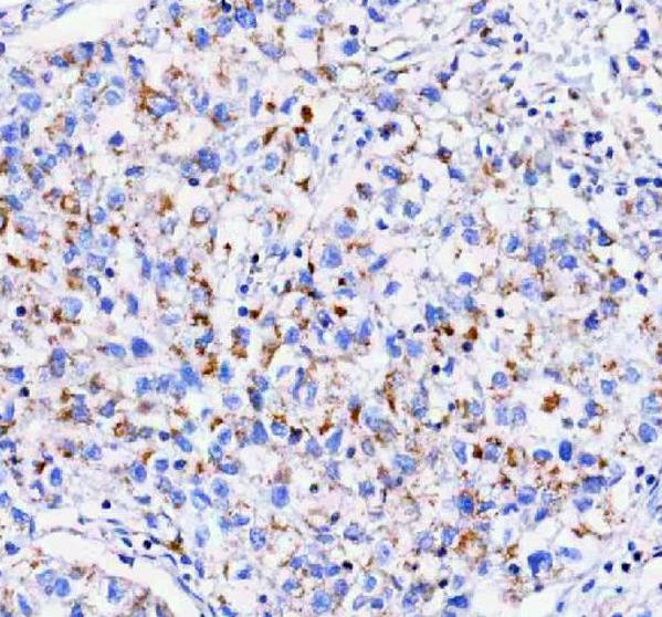
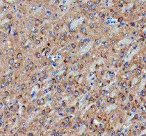
ARG44438 anti-IDUA antibody IHC-P image
Immunohistochemistry: Human lung cancer stained with ARG44438 anti-IDUA antibody at 2 μg/mL dilution.
-

ARG44438 anti-IDUA antibody IHC-P image
Immunohistochemistry: Human lung cancer stained with ARG44438 anti-IDUA antibody at 5 μg/mL dilution.
-
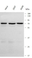
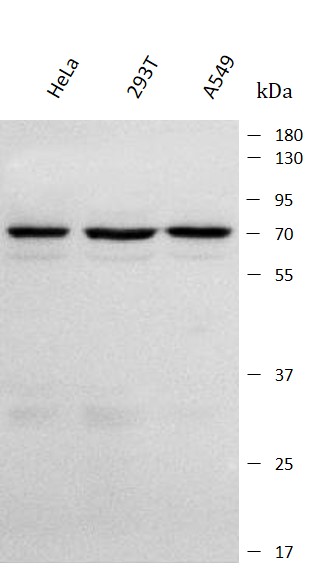
ARG44438 anti-IDUA antibody WB image
Western blot: HeLa, 293T and A549 stained with ARG44438 anti-IDUA antibody at 0.5 μg/mL dilution.
-
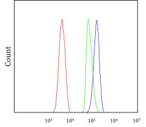
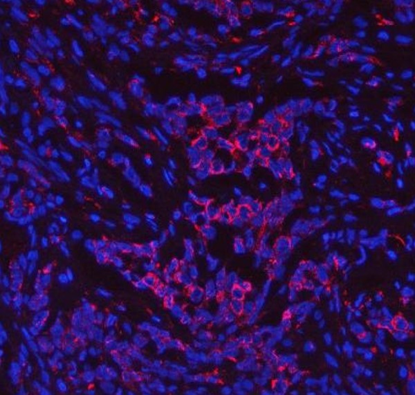
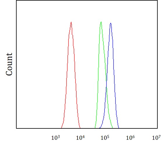
ARG44438 anti-IDUA antibody FACS image
Flow Cytometry: SH-SY5Y stained with ARG44438 anti-IDUA antibody at 1 μg/10^6 cells dilution.
-
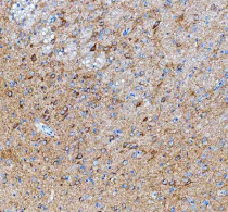
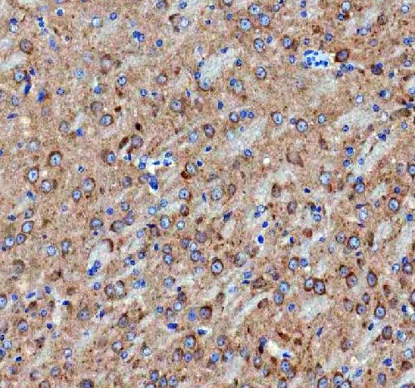
ARG44438 anti-IDUA antibody IHC-P image
Immunohistochemistry: Rat brain stained with ARG44438 anti-IDUA antibody at 2 μg/mL dilution.
-
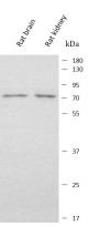
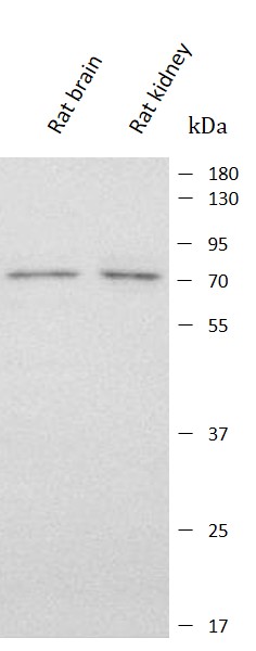
ARG44438 anti-IDUA antibody WB image
Western blot: Rat brain and Rat kidney stained with ARG44438 anti-IDUA antibody at 0.5 μg/mL dilution.
-
ARG44438 anti-IDUA antibody IHC-P image
Immunohistochemistry: Mouse brain stained with ARG44438 anti-IDUA antibody at 2 μg/mL dilution.
-

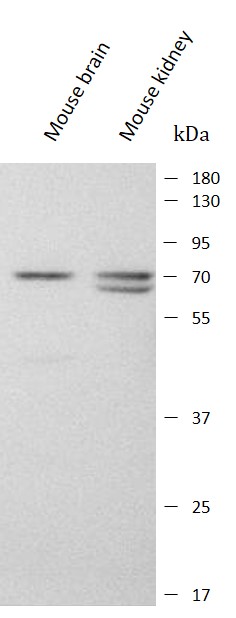
ARG44438 anti-IDUA antibody WB image
Western blot: Mouse brain and Mouse kidney stained with ARG44438 anti-IDUA antibody at 0.5 μg/mL dilution.