ARG42179
anti-IDH3G antibody
anti-IDH3G antibody for IHC-Formalin-fixed paraffin-embedded sections,Western blot and Human,Rat,Pig
Overview
| Product Description | Goat Polyclonal antibody recognizes IDH3G |
|---|---|
| Tested Reactivity | Hu, Rat, Pig |
| Predict Reactivity | Ms, Cow, Dog |
| Tested Application | IHC-P, WB |
| Host | Goat |
| Clonality | Polyclonal |
| Isotype | IgG |
| Target Name | IDH3G |
| Antigen Species | Human |
| Immunogen | Synthetic peptide around the internal region of Human IDH3G. (C-DHLKLHSYATSIRK) (NP_004126.1; NP_777358.1) |
| Conjugation | Un-conjugated |
| Alternate Names | NAD; Isocitric dehydrogenase subunit gamma; +; Isocitrate dehydrogenase [NAD] subunit gamma, mitochondrial; EC 1.1.1.41; H-IDHG |
Application Instructions
| Application Suggestion |
|
||||||
|---|---|---|---|---|---|---|---|
| Application Note | WB: Recommend incubate at RT for 1h. IHC-P: Antigen Retrieval: Steam tissue section in Citrate buffer (pH 6.0). * The dilutions indicate recommended starting dilutions and the optimal dilutions or concentrations should be determined by the scientist. |
||||||
| Positive Control | MCF7, Rat heart and Pig heart | ||||||
| Observed Size | ~ 40 kDa |
Properties
| Form | Liquid |
|---|---|
| Purification | Ammonium sulphate precipitation followed by affinity purification with immunogen. |
| Buffer | Tris saline (pH 7.3), 0.02% Sodium azide and 0.5% BSA. |
| Preservative | 0.02% Sodium azide |
| Stabilizer | 0.5% BSA |
| Concentration | 0.5 mg/ml |
| Storage Instruction | For continuous use, store undiluted antibody at 2-8°C for up to a week. For long-term storage, aliquot and store at -20°C or below. Storage in frost free freezers is not recommended. Avoid repeated freeze/thaw cycles. Suggest spin the vial prior to opening. The antibody solution should be gently mixed before use. |
| Note | For laboratory research only, not for drug, diagnostic or other use. |
Bioinformation
| Database Links |
Swiss-port # P51553 Human Isocitrate dehydrogenase [NAD] subunit gamma, mitochondrial |
|---|---|
| Gene Symbol | IDH3G |
| Gene Full Name | isocitrate dehydrogenase 3 (NAD+) gamma |
| Background | Isocitrate dehydrogenases catalyze the oxidative decarboxylation of isocitrate to 2-oxoglutarate. These enzymes belong to two distinct subclasses, one of which utilizes NAD(+) as the electron acceptor and the other NADP(+). Five isocitrate dehydrogenases have been reported: three NAD(+)-dependent isocitrate dehydrogenases, which localize to the mitochondrial matrix, and two NADP(+)-dependent isocitrate dehydrogenases, one of which is mitochondrial and the other predominantly cytosolic. NAD(+)-dependent isocitrate dehydrogenases catalyze the allosterically regulated rate-limiting step of the tricarboxylic acid cycle. Each isozyme is a heterotetramer that is composed of two alpha subunits, one beta subunit, and one gamma subunit. The protein encoded by this gene is the gamma subunit of one isozyme of NAD(+)-dependent isocitrate dehydrogenase. This gene is a candidate gene for periventricular heterotopia. Several alternatively spliced transcript variants of this gene have been described, but only some of their full length natures have been determined. [provided by RefSeq, Jul 2008] |
| Function | Regulatory subunit which plays a role in the allosteric regulation of the enzyme catalyzing the decarboxylation of isocitrate (ICT) into alpha-ketoglutarate. The heterodimer composed of the alpha (IDH3A) and beta (IDH3B) subunits and the heterodimer composed of the alpha (IDH3A) and gamma (IDH3G) subunits, have considerable basal activity but the full activity of the heterotetramer (containing two subunits of IDH3A, one of IDH3B and one of IDH3G) requires the assembly and cooperative function of both heterodimers. [UniProt] |
| Cellular Localization | Mitochondrion. [UniProt] |
| Calculated MW | 43 kDa |
Images (3) Click the Picture to Zoom In
-
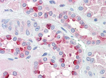
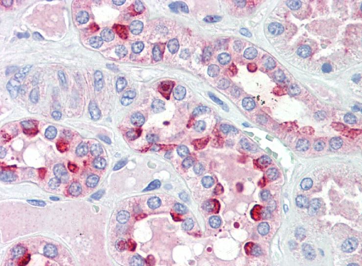
ARG42179 anti-IDH3G antibody IHC-P image
Immunohistochemistry: Paraffin-embedded Human kidney tissue. Antigen Retrieval: Steam tissue section in Citrate buffer (pH 6.0). The tissue section was stained with ARG42179 anti-IDH3G antibody at 5 µg/ml dilution followed by AP-staining.
-
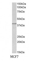
ARG42179 anti-IDH3G antibody WB image
Western blot: 35 µg of MCF7 cell lysate (in RIPA buffer) stained with ARG42179 anti-IDH3G antibody at 0.5 µg/ml dilution and incubated at RT for 1 hour.
-
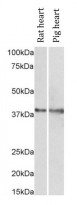
ARG42179 anti-IDH3G antibody WB image
Western blot: 35 µg of Rat heart and Pig heart lysate (in RIPA buffer) stained with ARG42179 anti-IDH3G antibody at 1 µg/ml dilution and incubated at RT for 1 hour.