ARG42154
anti-IDH3A antibody
anti-IDH3A antibody for ICC/IF,IHC-Formalin-fixed paraffin-embedded sections,Immunoprecipitation,Western blot and Human,Mouse
Overview
| Product Description | Rabbit Polyclonal antibody recognizes IDH3A |
|---|---|
| Tested Reactivity | Hu, Ms |
| Predict Reactivity | Cow, Rat, Dog, Gpig, Hrs, Rb, Zfsh |
| Tested Application | ICC/IF, IHC-P, IP, WB |
| Host | Rabbit |
| Clonality | Polyclonal |
| Isotype | IgG |
| Target Name | IDH3A |
| Antigen Species | Human |
| Immunogen | Synthetic peptide around the N-terminal region of Human IDH3A. (within the following region: MKIFD AAKAP IQWEE RNVTA IQGPG GKWMI PSEAK ESMDK NKMGL KGPLK) |
| Conjugation | Un-conjugated |
| Alternate Names | Isocitrate dehydrogenase [NAD] subunit alpha, mitochondrial; EC 1.1.1.41; NAD; Isocitric dehydrogenase subunit alpha; + |
Application Instructions
| Predict Reactivity Note | Predicted Homology Based on Immunogen Sequence: Cow: 100%; Dog: 100%; Guinea pig: 100%; Horse: 100%; Rabbit: 100%; Rat: 100%; Zebrafish: 92% | ||||||||||
|---|---|---|---|---|---|---|---|---|---|---|---|
| Application Suggestion |
|
||||||||||
| Application Note | * The dilutions indicate recommended starting dilutions and the optimal dilutions or concentrations should be determined by the scientist. | ||||||||||
| Positive Control | HepG2 | ||||||||||
| Observed Size | ~ 38 - 40 kDa |
Properties
| Form | Liquid |
|---|---|
| Purification | Purification with Protein A. |
| Buffer | PBS, 0.09% (w/v) Sodium azide and 2% Sucrose. |
| Preservative | 0.09% (w/v) Sodium azide |
| Stabilizer | 2% Sucrose |
| Concentration | Batch dependent: 0.5 - 1 mg/ml |
| Storage Instruction | For continuous use, store undiluted antibody at 2-8°C for up to a week. For long-term storage, aliquot and store at -20°C or below. Storage in frost free freezers is not recommended. Avoid repeated freeze/thaw cycles. Suggest spin the vial prior to opening. The antibody solution should be gently mixed before use. |
| Note | For laboratory research only, not for drug, diagnostic or other use. |
Bioinformation
| Database Links |
Swiss-port # P50213 Human Isocitrate dehydrogenase [NAD] subunit alpha, mitochondrial Swiss-port # Q9D6R2 Mouse Isocitrate dehydrogenase [NAD] subunit alpha, mitochondrial |
|---|---|
| Gene Symbol | IDH3A |
| Gene Full Name | isocitrate dehydrogenase 3 (NAD+) alpha |
| Background | Isocitrate dehydrogenases catalyze the oxidative decarboxylation of isocitrate to 2-oxoglutarate. These enzymes belong to two distinct subclasses, one of which utilizes NAD(+) as the electron acceptor and the other NADP(+). Five isocitrate dehydrogenases have been reported: three NAD(+)-dependent isocitrate dehydrogenases, which localize to the mitochondrial matrix, and two NADP(+)-dependent isocitrate dehydrogenases, one of which is mitochondrial and the other predominantly cytosolic. NAD(+)-dependent isocitrate dehydrogenases catalyze the allosterically regulated rate-limiting step of the tricarboxylic acid cycle. Each isozyme is a heterotetramer that is composed of two alpha subunits, one beta subunit, and one gamma subunit. The protein encoded by this gene is the alpha subunit of one isozyme of NAD(+)-dependent isocitrate dehydrogenase. [provided by RefSeq, Jul 2008] |
| Function | Catalytic subunit of the enzyme which catalyzes the decarboxylation of isocitrate (ICT) into alpha-ketoglutarate. The heterodimer composed of the alpha (IDH3A) and beta (IDH3B) subunits and the heterodimer composed of the alpha (IDH3A) and gamma (IDH3G) subunits, have considerable basal activity but the full activity of the heterotetramer (containing two subunits of IDH3A, one of IDH3B and one of IDH3G) requires the assembly and cooperative function of both heterodimers. [UniProt] |
| Cellular Localization | Mitochondrion. [UniProt] |
| Calculated MW | 40 kDa |
Images (4) Click the Picture to Zoom In
-
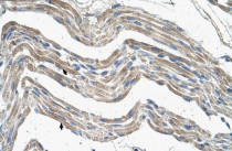
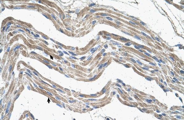
ARG42154 anti-IDH3A antibody IHC-P image
Immunohistochemistry: Paraffin-embedded Human skeletal muscle tissue stained with ARG42154 anti-IDH3A antibody at 4 - 8 µg/ml dilution.
-

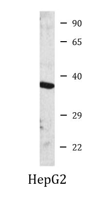
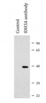
ARG42154 anti-IDH3A antibody WB image
Western blot: HepG2 cell lysate stained with ARG42154 anti-IDH3A antibody at 1 µg/ml dilution.
-
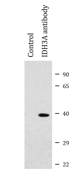
ARG42154 anti-IDH3A antibody IP image
Immunoprecipitation: 2 mg of HEK293 whole cell lysate were immunoprecipitated (1:200) and stained with ARG42154 anti-IDH3A antibody at 1:1000 dilution.
-
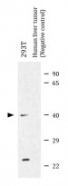
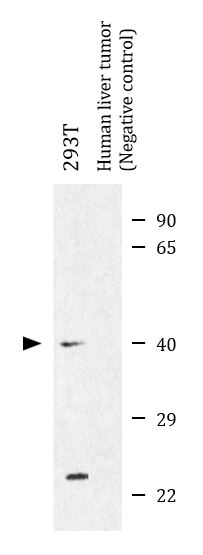
ARG42154 anti-IDH3A antibody WB image
Western blot: 25 µg of 293T and Human liver tumor (Negative control) lysates stained with ARG42154 anti-IDH3A antibody at 5 µg/ml dilution.