ARG42681
anti-IDE / Insulysin antibody
anti-IDE / Insulysin antibody for Flow cytometry,IHC-Formalin-fixed paraffin-embedded sections,Western blot and Human,Mouse,Rat
Overview
| Product Description | Rabbit Polyclonal antibody recognizes IDE / Insulysin |
|---|---|
| Tested Reactivity | Hu, Ms, Rat |
| Tested Application | FACS, IHC-P, WB |
| Host | Rabbit |
| Clonality | Polyclonal |
| Isotype | IgG |
| Target Name | IDE / Insulysin |
| Antigen Species | Human |
| Immunogen | Recombinant protein corresponding to F485-K756 of Human IDE / Insulysin. |
| Conjugation | Un-conjugated |
| Alternate Names | Insulysin; Abeta-degrading protease; Insulinase; EC 3.4.24.56; Insulin protease; INSULYSIN; Insulin-degrading enzyme |
Application Instructions
| Application Suggestion |
|
||||||||
|---|---|---|---|---|---|---|---|---|---|
| Application Note | * The dilutions indicate recommended starting dilutions and the optimal dilutions or concentrations should be determined by the scientist. | ||||||||
| Observed Size | ~ 120 kDa |
Properties
| Form | Liquid |
|---|---|
| Purification | Affinity purification with immunogen. |
| Buffer | 0.2% Na2HPO4, 0.9% NaCl, 0.05% Sodium azide and 4% Trehalose. |
| Preservative | 0.05% Sodium azide |
| Stabilizer | 4% Trehalose |
| Concentration | 0.5 mg/ml |
| Storage Instruction | For continuous use, store undiluted antibody at 2-8°C for up to a week. For long-term storage, aliquot and store at -20°C or below. Storage in frost free freezers is not recommended. Avoid repeated freeze/thaw cycles. Suggest spin the vial prior to opening. The antibody solution should be gently mixed before use. |
| Note | For laboratory research only, not for drug, diagnostic or other use. |
Bioinformation
| Database Links | |
|---|---|
| Gene Symbol | IDE |
| Gene Full Name | insulin-degrading enzyme |
| Background | This gene encodes a zinc metallopeptidase that degrades intracellular insulin, and thereby terminates insulins activity, as well as participating in intercellular peptide signalling by degrading diverse peptides such as glucagon, amylin, bradykinin, and kallidin. The preferential affinity of this enzyme for insulin results in insulin-mediated inhibition of the degradation of other peptides such as beta-amyloid. Deficiencies in this protein's function are associated with Alzheimer's disease and type 2 diabetes mellitus but mutations in this gene have not been shown to be causitive for these diseases. This protein localizes primarily to the cytoplasm but in some cell types localizes to the extracellular space, cell membrane, peroxisome, and mitochondrion. Alternative splicing results in multiple transcript variants encoding distinct isoforms. Additional transcript variants have been described but have not been experimentally verified. [provided by RefSeq, Sep 2009] |
| Function | Plays a role in the cellular breakdown of insulin, APP peptides, IAPP peptides, glucagon, bradykinin, kallidin and other peptides, and thereby plays a role in intercellular peptide signaling (PubMed:2293021, PubMed:10684867, PubMed:26968463, PubMed:17051221, PubMed:17613531, PubMed:18986166, PubMed:19321446, PubMed:23922390, PubMed:24847884, PubMed:26394692, PubMed:29596046). Substrate binding induces important conformation changes, making it possible to bind and degrade larger substrates, such as insulin (PubMed:23922390, PubMed:26394692, PubMed:29596046). Contributes to the regulation of peptide hormone signaling cascades and regulation of blood glucose homeostasis via its role in the degradation of insulin, glucagon and IAPP (By similarity). Plays a role in the degradation and clearance of APP-derived amyloidogenic peptides that are secreted by neurons and microglia (PubMed:9830016, PubMed:26394692) (Probable). Involved in antigen processing. Produces both the N terminus and the C terminus of MAGEA3-derived antigenic peptide (EVDPIGHLY) that is presented to cytotoxic T lymphocytes by MHC class I. (Microbial infection) The membrane-associated isoform acts as an entry receptor for varicella-zoster virus (VZV). [UniProt] |
| Cellular Localization | Cytoplasm. Cell membrane. Secreted. Note=Present at the cell surface of neuron cells. The membrane-associated isoform is approximately 5 kDa larger than the known cytosolic isoform. [UniProt] |
| Calculated MW | 118 kDa |
| PTM | The N-terminus is blocked. [UniProt] |
Images (5) Click the Picture to Zoom In
-
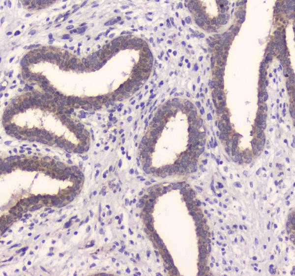
ARG42681 anti-IDE / Insulysin antibody IHC-P image
Immunohistochemistry: Paraffin-embedded Human mammary cancer tissue. Antigen Retrieval: Heat mediation was performed in Citrate buffer (pH 6.0) for 20 min. The tissue section was blocked with 10% goat serum. The tissue section was then stained with ARG42681 anti-IDE / Insulysin antibody at 1 µg/ml dilution, overnight at 4°C.
-

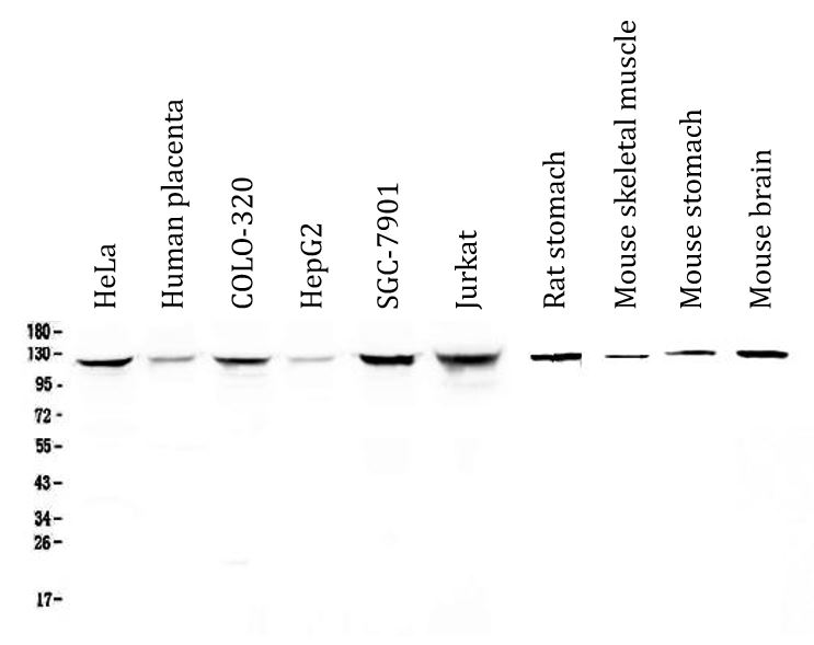
ARG42681 anti-IDE / Insulysin antibody WB image
Western blot: 50 µg of samples under reducing condition. HeLa, Human placenta, COLO-320, HepG2, SGC-7901, Jurkat, Rat stomach, Mouse skeletal muscle, Mouse stomach and Mouse brain lysates stained with ARG42681 anti-IDE / Insulysin antibody at 0.5 µg/ml dilution, overnight at 4°C.
-
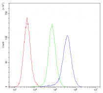
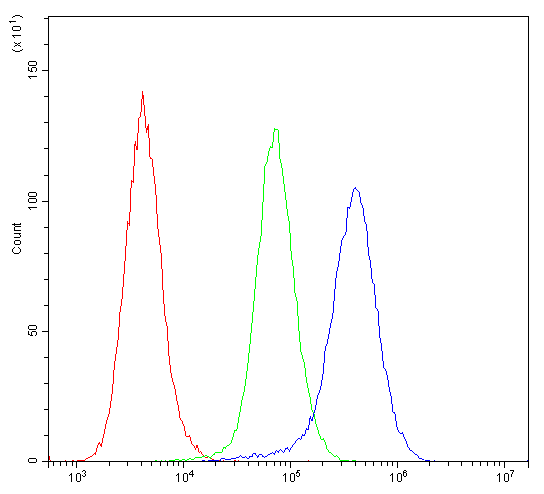
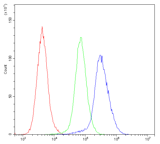
ARG42681 anti-IDE / Insulysin antibody FACS image
Flow Cytometry: HeLa cells were blocked with 10% normal goat serum and then stained with ARG42681 anti-IDE / Insulysin antibody (blue) at 1 µg/10^6 cells for 30 min at 20°C, followed by incubation with DyLight®488 labelled secondary antibody. Isotype control antibody (green) was rabbit IgG (1 µg/10^6 cells) used under the same conditions. Unlabelled sample (red) was also used as a control.
-

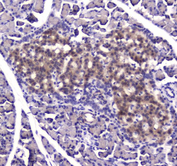
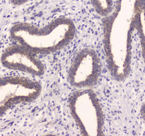
ARG42681 anti-IDE / Insulysin antibody IHC-P image
Immunohistochemistry: Paraffin-embedded Rat pancreas tissue. Antigen Retrieval: Heat mediation was performed in Citrate buffer (pH 6.0) for 20 min. The tissue section was blocked with 10% goat serum. The tissue section was then stained with ARG42681 anti-IDE / Insulysin antibody at 1 µg/ml dilution, overnight at 4°C.
-
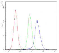
ARG42681 anti-IDE / Insulysin antibody FACS image
Flow Cytometry: CACO-2 cells were blocked with 10% normal goat serum and then stained with ARG42681 anti-IDE / Insulysin antibody (blue) at 1 µg/10^6 cells for 30 min at 20°C, followed by incubation with DyLight®488 labelled secondary antibody. Isotype control antibody (green) was rabbit IgG (1 µg/10^6 cells) used under the same conditions. Unlabelled sample (red) was also used as a control.