ARG40993
anti-Hsp 70 antibody
anti-Hsp 70 antibody for Flow cytometry,ICC/IF,IHC-Formalin-fixed paraffin-embedded sections,Western blot and Human,Mouse,Rat
Overview
| Product Description | Rabbit Polyclonal antibody recognizes Hsp 70 |
|---|---|
| Tested Reactivity | Hu, Ms, Rat |
| Predict Reactivity | Bov, Hrs, Mk |
| Tested Application | FACS, ICC/IF, IHC-P, WB |
| Host | Rabbit |
| Clonality | Polyclonal |
| Isotype | IgG |
| Target Name | Hsp 70 |
| Antigen Species | Human |
| Immunogen | Synthetic peptide corresponding to aa. 559-596 of Human Hsp 70 (KGKISEADKKKVLDKCQEVISWLDANTLAEKDEFEHKR). |
| Conjugation | Un-conjugated |
| Alternate Names | Heat shock 70 kDa protein 1A; HSPA1; HSP70I; Heat shock 70 kDa protein 1; HSP70-1A; HEL-S-103; HSP70.1; HSP72; HSP70-1 |
Application Instructions
| Application Suggestion |
|
||||||||||
|---|---|---|---|---|---|---|---|---|---|---|---|
| Application Note | * The dilutions indicate recommended starting dilutions and the optimal dilutions or concentrations should be determined by the scientist. |
Properties
| Form | Liquid |
|---|---|
| Purification | Affinity purification with immunogen. |
| Buffer | 0.2% Na2HPO4, 0.9% NaCl, 0.05% Sodium azide and 5% BSA. |
| Preservative | 0.05% Sodium azide |
| Stabilizer | 5% BSA |
| Concentration | 0.5 mg/ml |
| Storage Instruction | For continuous use, store undiluted antibody at 2-8°C for up to a week. For long-term storage, aliquot and store at -20°C or below. Storage in frost free freezers is not recommended. Avoid repeated freeze/thaw cycles. Suggest spin the vial prior to opening. The antibody solution should be gently mixed before use. |
| Note | For laboratory research only, not for drug, diagnostic or other use. |
Bioinformation
| Database Links | |
|---|---|
| Gene Symbol | HSPA1A |
| Gene Full Name | heat shock 70kDa protein 1A |
| Background | This intronless gene encodes a 70kDa heat shock protein which is a member of the heat shock protein 70 family. In conjuction with other heat shock proteins, this protein stabilizes existing proteins against aggregation and mediates the folding of newly translated proteins in the cytosol and in organelles. It is also involved in the ubiquitin-proteasome pathway through interaction with the AU-rich element RNA-binding protein 1. The gene is located in the major histocompatibility complex class III region, in a cluster with two closely related genes which encode similar proteins. [provided by RefSeq, Jul 2008] |
| Function | In cooperation with other chaperones, Hsp70s stabilize preexistent proteins against aggregation and mediate the folding of newly translated polypeptides in the cytosol as well as within organelles. These chaperones participate in all these processes through their ability to recognize nonnative conformations of other proteins. They bind extended peptide segments with a net hydrophobic character exposed by polypeptides during translation and membrane translocation, or following stress-induced damage. In case of rotavirus A infection, serves as a post-attachment receptor for the virus to facilitate entry into the cell. Essential for STUB1-mediated ubiquitination and degradation of FOXP3 in regulatory T-cells (Treg) during inflammation. [UniProt] |
| Cellular Localization | Cytoplasm. Nucleus. Cytoplasm, cytoskeleton, microtubule organizing center, centrosome. Note=Localized in cytoplasmic mRNP granules containing untranslated mRNAs. [UniProt] |
| Calculated MW | 70 kDa |
| PTM | In response to cellular stress, acetylated at Lys-77 by NA110 and then gradually deacetylated by HDAC4 at later stages. Acetylation enhances its chaperone activity and also determines whether it will function as a chaperone for protein refolding or degradation by controlling its binding to co-chaperones HOPX and STUB1. The acetylated form and the non-acetylated form bind to HOPX and STUB1 respectively. Acetylation also protects cells against various types of cellular stress. [UniProt] |
Images (6) Click the Picture to Zoom In
-
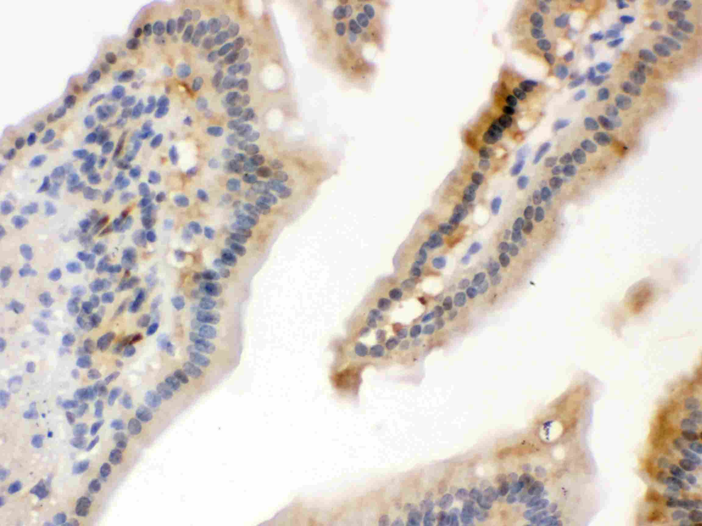
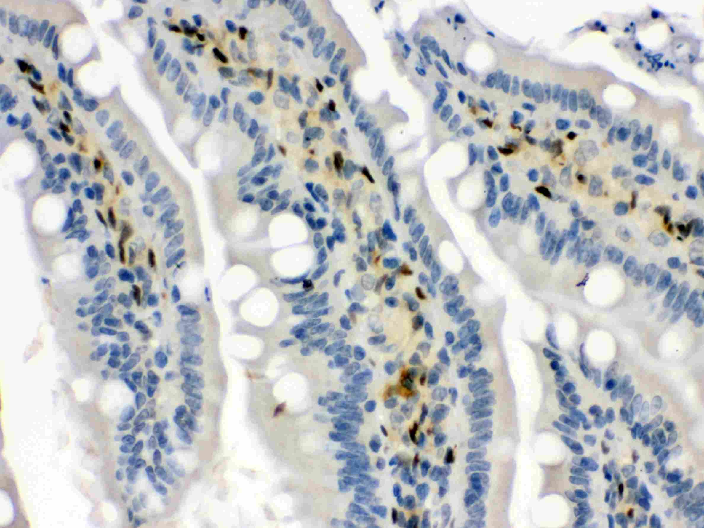
ARG40993 anti-Hsp 70 antibody IHC-P image
Immunohistochemistry: Paraffin-embedded Mouse intestine tissue stained with ARG40993 anti-Hsp 70 antibody.
-

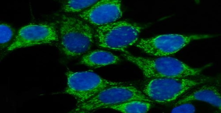
ARG40993 anti-Hsp 70 antibody ICC/IF image
Immunofluorescence: MCF-7 stained with ARG40993 anti-Hsp 70 antibody at 5 μg/ml dilution.
-
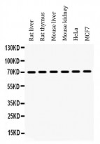
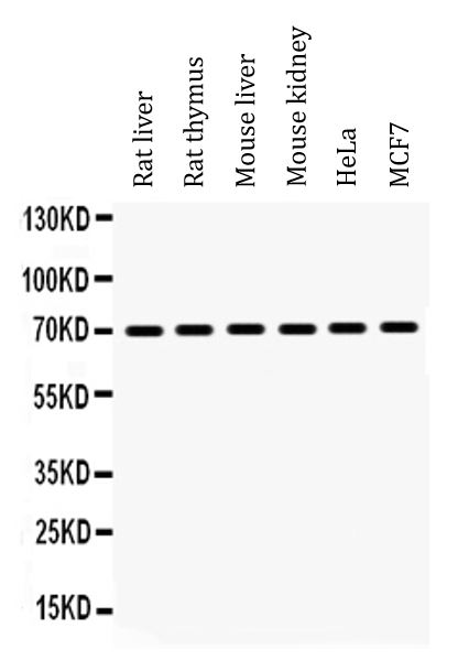
ARG40993 anti-Hsp 70 antibody WB image
Western blot: 50 µg of Rat liver, 50 µg of Rat thymus, 50 µg of Mouse liver, 50 µg of Mouse kidney, 40 µg of HeLa and 40 µg of MCF7 whole cell lysates stained with ARG40993 anti-Hsp 70 antibody at 0.5 µg/ml dilution.
-
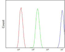
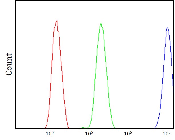
ARG40993 anti-Hsp 70 antibody FACS image
Flow Cytometry: Hela stained with ARG40993 anti-Hsp 70 antibody at 1-3 μg/1x10^6 cells dilution.
-
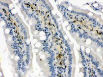
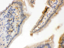
ARG40993 anti-Hsp 70 antibody IHC-P image
Immunohistochemistry: Paraffin-embedded Rat intestine tissue stained with ARG40993 anti-Hsp 70 antibody.
-
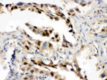
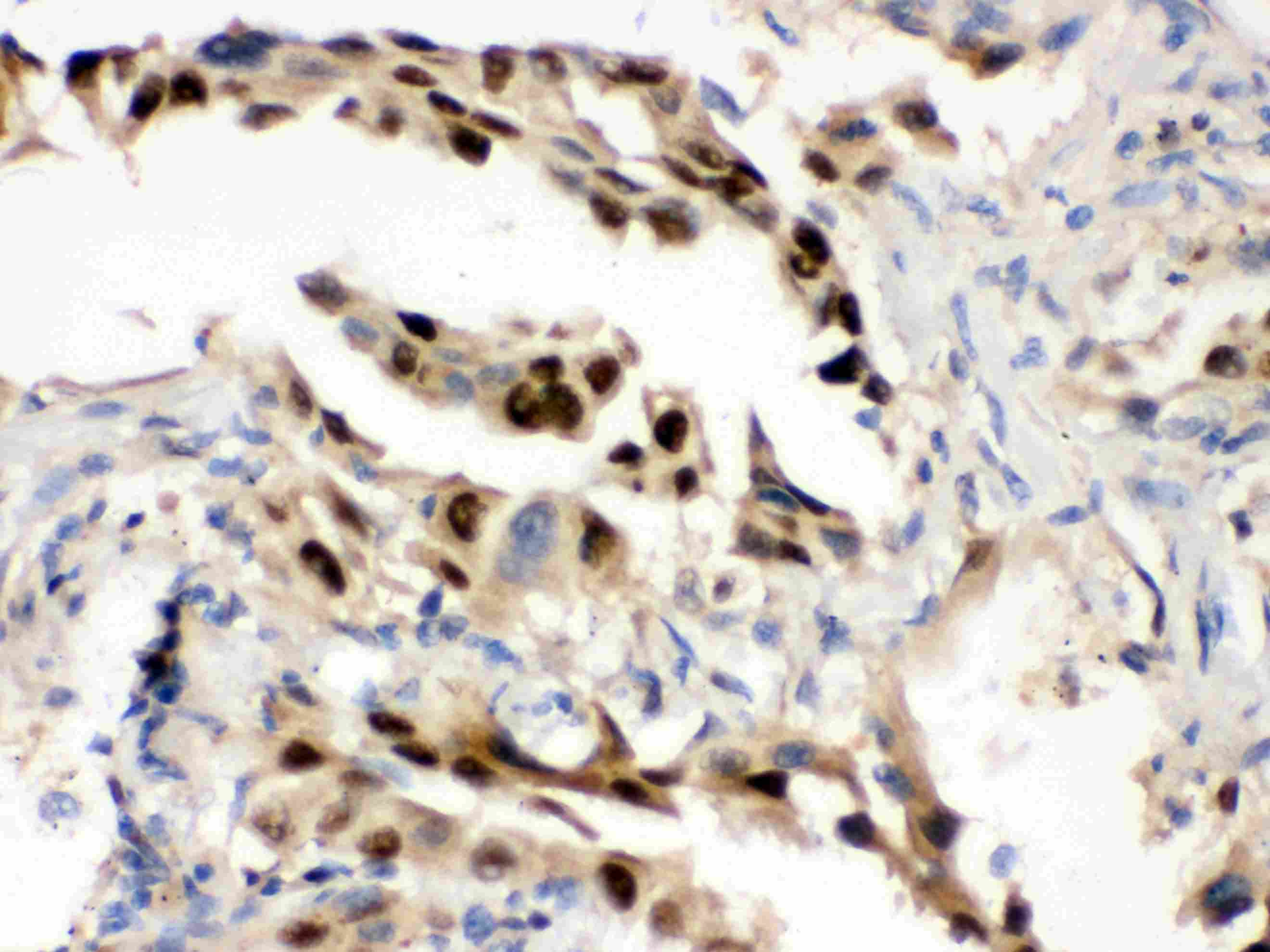
ARG40993 anti-Hsp 70 antibody IHC-P image
Immunohistochemistry: Paraffin-embedded Human lung cancer tissue stained with ARG40993 anti-Hsp 70 antibody.