ARG55373
anti-Hsc 70 antibody
anti-Hsc 70 antibody for Flow cytometry,ICC/IF,IHC-Formalin-fixed paraffin-embedded sections,Western blot and Human,Rat
Signaling Transduction antibody
Overview
| Product Description | Rabbit Polyclonal antibody recognizes Hsc 70 |
|---|---|
| Tested Reactivity | Hu, Rat |
| Predict Reactivity | Ms, Bov, Chk, Hm |
| Tested Application | FACS, ICC/IF, IHC-P, WB |
| Host | Rabbit |
| Clonality | Polyclonal |
| Isotype | IgG |
| Target Name | Hsc 70 |
| Antigen Species | Human |
| Immunogen | KLH-conjugated synthetic peptide corresponding to aa. 82-110 (N-terminus) of Human Hsc 70. |
| Conjugation | Un-conjugated |
| Alternate Names | NIP71; HEL-33; HSPA10; LAP-1; HSC70; HSC71; LAP1; Lipopolysaccharide-associated protein 1; Heat shock 70 kDa protein 8; HSC54; HEL-S-72p; Heat shock cognate 71 kDa protein; HSP71; HSP73; LPS-associated protein 1 |
Application Instructions
| Application Suggestion |
|
||||||||||
|---|---|---|---|---|---|---|---|---|---|---|---|
| Application Note | * The dilutions indicate recommended starting dilutions and the optimal dilutions or concentrations should be determined by the scientist. | ||||||||||
| Positive Control | A431 |
Properties
| Form | Liquid |
|---|---|
| Purification | This antibody is prepared by Saturated Ammonium Sulfate (SAS) precipitation followed by dialysis against PBS. |
| Buffer | PBS and 0.09% (W/V) Sodium azide |
| Preservative | 0.09% (W/V) Sodium azide |
| Storage Instruction | For continuous use, store undiluted antibody at 2-8°C for up to a week. For long-term storage, aliquot and store at -20°C or below. Storage in frost free freezers is not recommended. Avoid repeated freeze/thaw cycles. Suggest spin the vial prior to opening. The antibody solution should be gently mixed before use. |
| Note | For laboratory research only, not for drug, diagnostic or other use. |
Bioinformation
| Database Links | |
|---|---|
| Gene Symbol | HSPA8 |
| Gene Full Name | heat shock 70kDa protein 8 |
| Background | This gene encodes a member of the heat shock protein 70 family, which contains both heat-inducible and constitutively expressed members. This protein belongs to the latter group, which are also referred to as heat-shock cognate proteins. It functions as a chaperone, and binds to nascent polypeptides to facilitate correct folding. It also functions as an ATPase in the disassembly of clathrin-coated vesicles during transport of membrane components through the cell. Alternatively spliced transcript variants encoding different isoforms have been found for this gene. [provided by RefSeq, Aug 2011] |
| Function | Acts as a repressor of transcriptional activation. Inhibits the transcriptional coactivator activity of CITED1 on Smad-mediated transcription. Chaperone. Component of the PRP19-CDC5L complex that forms an integral part of the spliceosome and is required for activating pre-mRNA splicing. May have a scaffolding role in the spliceosome assembly as it contacts all other components of the core complex. Binds bacterial lipopolysaccharide (LPS) et mediates LPS-induced inflammatory response, including TNF secretion by monocytes. Participates in the ER-associated degradation (ERAD) quality control pathway in conjunction with J domain-containing co-chaperones and the E3 ligase CHIP. [UniProt] |
| Cellular Localization | Cytoplasm. Melanosome. Nucleus, nucleolus. Cell membrane. Note=Localized in cytoplasmic mRNP granules containing untranslated mRNAs. Translocates rapidly from the cytoplasm to the nuclei, and especially to the nucleoli, upon heat shock |
| Research Area | Signaling Transduction antibody |
| Calculated MW | 71 kDa |
| PTM | Acetylated. ISGylated. Trimethylation at Lys-561 reduces fibrillar SNCA binding. |
Images (4) Click the Picture to Zoom In
-
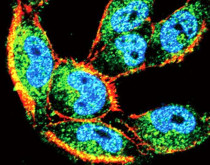
ARG55373 anti-Hsc 70 antibody ICC/IF image
Immunofluorescence: A2058 cells stained with ARG55373 anti-Hsc 70 antibody (green). Actin filaments have been labeled with Alexa Fluor 555 phalloidin (red). DAPI (blue) for nuclear staining.
-
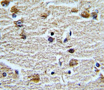
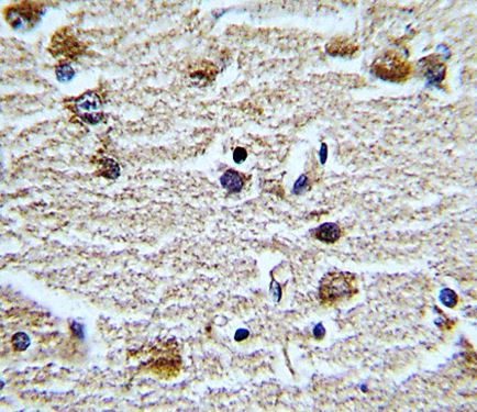
ARG55373 anti-Hsc 70 antibody IHC-P image
Immunohistochemistry: Formalin-fixed and paraffin-embedded Human brain tissue stained with ARG55373 anti-Hsc 70 antibody.
-
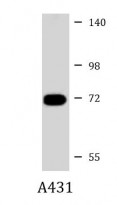
ARG55373 anti-Hsc 70 antibody WB image
Western blot: 35 µg of A431 cell lysate stained with ARG55373 anti-Hsc 70 antibody at 1:1000 dilution.
-

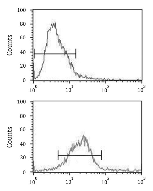
ARG55373 anti-Hsc 70 antibody FACS image
Flow Cytometry: HeLa cells stained with ARG55373 anti-Hsc 70 antibody (bottom histogram) or without primary antibody control (top histogram), followed by incubation with FITC labelled secondary antibody.