ARG59625
anti-Hck antibody
anti-Hck antibody for ICC/IF,Western blot and Human
Overview
| Product Description | Mouse Monoclonal antibody recognizes Hck |
|---|---|
| Tested Reactivity | Hu |
| Tested Application | ICC/IF, WB |
| Host | Mouse |
| Clonality | Monoclonal |
| Clone | 1508CT602.13.1 |
| Isotype | IgG1, kappa |
| Target Name | Hck |
| Antigen Species | Human |
| Immunogen | Recombinant protein of Human Hck. |
| Conjugation | Un-conjugated |
| Alternate Names | JTK9; Hematopoietic cell kinase; p59-HCK/p60-HCK; p61Hck; Hemopoietic cell kinase; Tyrosine-protein kinase HCK; p59Hck; EC 2.7.10.2 |
Application Instructions
| Application Suggestion |
|
||||||
|---|---|---|---|---|---|---|---|
| Application Note | * The dilutions indicate recommended starting dilutions and the optimal dilutions or concentrations should be determined by the scientist. | ||||||
| Positive Control | THP-1 |
Properties
| Form | Liquid |
|---|---|
| Purification | Purification with Protein G. |
| Buffer | PBS and 0.09% (W/V) Sodium azide. |
| Preservative | 0.09% (W/V) Sodium azide |
| Storage Instruction | For continuous use, store undiluted antibody at 2-8°C for up to a week. For long-term storage, aliquot and store at -20°C or below. Storage in frost free freezers is not recommended. Avoid repeated freeze/thaw cycles. Suggest spin the vial prior to opening. The antibody solution should be gently mixed before use. |
| Note | For laboratory research only, not for drug, diagnostic or other use. |
Bioinformation
| Database Links | |
|---|---|
| Gene Symbol | HCK |
| Gene Full Name | HCK proto-oncogene, Src family tyrosine kinase |
| Background | The protein encoded by this gene is a member of the Src family of tyrosine kinases. This protein is primarily hemopoietic, particularly in cells of the myeloid and B-lymphoid lineages. It may help couple the Fc receptor to the activation of the respiratory burst. In addition, it may play a role in neutrophil migration and in the degranulation of neutrophils. Multiple isoforms with different subcellular distributions are produced due to both alternative splicing and the use of alternative translation initiation codons, including a non-AUG (CUG) codon. [provided by RefSeq, Feb 2010] |
| Function | Non-receptor tyrosine-protein kinase found in hematopoietic cells that transmits signals from cell surface receptors and plays an important role in the regulation of innate immune responses, including neutrophil, monocyte, macrophage and mast cell functions, phagocytosis, cell survival and proliferation, cell adhesion and migration. Acts downstream of receptors that bind the Fc region of immunoglobulins, such as FCGR1A and FCGR2A, but also CSF3R, PLAUR, the receptors for IFNG, IL2, IL6 and IL8, and integrins, such as ITGB1 and ITGB2. During the phagocytic process, mediates mobilization of secretory lysosomes, degranulation, and activation of NADPH oxidase to bring about the respiratory burst. Plays a role in the release of inflammatory molecules. Promotes reorganization of the actin cytoskeleton and actin polymerization, formation of podosomes and cell protrusions. Inhibits TP73-mediated transcription activation and TP73-mediated apoptosis. Phosphorylates CBL in response to activation of immunoglobulin gamma Fc region receptors. Phosphorylates ADAM15, BCR, ELMO1, FCGR2A, GAB1, GAB2, RAPGEF1, STAT5B, TP73, VAV1 and WAS. [UniProt] |
| Cellular Localization | Isoform 1: Lysosome. Membrane; Lipid-anchor. Cell projection, podosome membrane; Lipid-anchor. Cytoplasm, cytosol. Isoform 2: Cell membrane; Lipid-anchor. Membrane, caveola; Lipid-anchor. Cell junction, focal adhesion. Cytoplasm, cytoskeleton. Golgi apparatus. Cytoplasmic vesicle. Lysosome. Nucleus. [UniProt] |
| Calculated MW | 60 kDa |
| PTM | Phosphorylated on several tyrosine residues. Autophosphorylated. Becomes rapidly phosphorylated upon activation of the immunoglobulin receptors FCGR1A and FCGR2A. Phosphorylation by the BCR-ABL fusion protein mediates activation of HCK. Phosphorylation at Tyr-411 increases kinase activity. Phosphorylation at Tyr-522 inhibits kinase activity. Kinase activity is not required for phosphorylation at Tyr-522, suggesting that this site is a target of other kinases. Ubiquitinated by CBL, leading to its degradation via the proteasome. Isoform 2 palmitoylation at position 2 requires prior myristoylation. Palmitoylation at position 3 is required for caveolar localization of isoform 2. [UniProt] |
Images (2) Click the Picture to Zoom In
-

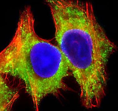
ARG59625 anti-Hck antibody ICC/IF image
Immunofluorescence: 4% paraformaldehyde-fixed, 0.1% Triton X-100 permeabilized HepG2 cells stained with ARG59625 anti-Hck antibody (green) at 1:25 dilution. Cytoplasmic actin is detected with Dylight® 554 Phalloidin (red) at 1:100 dilution. DAPI (blue) for nuclear staining.
-

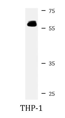
ARG59625 anti-Hck antibody WB image
Western blot: 20 µg of THP-1 cell lysate stained with ARG59625 anti-Hck antibody at 1:2000 - 1:4000 dilution.