ARG59688
anti-HSPA2 antibody [4A4]
anti-HSPA2 antibody [4A4] for Flow cytometry,ICC/IF,IHC-Formalin-fixed paraffin-embedded sections,Western blot and Human,Mouse,Rat
Overview
| Product Description | Mouse Monoclonal antibody [4A4] recognizes HSPA2 |
|---|---|
| Tested Reactivity | Hu, Ms, Rat |
| Tested Application | FACS, ICC/IF, IHC-P, WB |
| Host | Mouse |
| Clonality | Monoclonal |
| Clone | 4A4 |
| Isotype | IgG1 |
| Target Name | HSPA2 |
| Antigen Species | Human |
| Immunogen | Synthetic peptide corresponding to aa. 564-598 of Human HSPA2. (KISEQDKNKILDKCQEVINWLDRNQMAEKDEYEHK) |
| Conjugation | Un-conjugated |
| Alternate Names | Heat shock 70 kDa protein 2; Heat shock-related 70 kDa protein 2; HSP70-2; HSP70-3 |
Application Instructions
| Application Suggestion |
|
||||||||||
|---|---|---|---|---|---|---|---|---|---|---|---|
| Application Note | IHC-P: Antigen Retrieval: Heat mediation was performed in Citrate buffer (pH 6.0) for 20 min. * The dilutions indicate recommended starting dilutions and the optimal dilutions or concentrations should be determined by the scientist. |
Properties
| Form | Liquid |
|---|---|
| Purification | Affinity purification with immunogen. |
| Buffer | 0.9% NaCl, 0.2% Na2HPO4, 0.05% Sodium azide and 4% Trehalose. |
| Preservative | 0.05% Sodium azide |
| Stabilizer | 4% Trehalose |
| Concentration | 0.5 mg/ml |
| Storage Instruction | For continuous use, store undiluted antibody at 2-8°C for up to a week. For long-term storage, aliquot and store at -20°C or below. Storage in frost free freezers is not recommended. Avoid repeated freeze/thaw cycles. Suggest spin the vial prior to opening. The antibody solution should be gently mixed before use. |
| Note | For laboratory research only, not for drug, diagnostic or other use. |
Bioinformation
| Database Links | |
|---|---|
| Gene Symbol | HSPA2 |
| Gene Full Name | heat shock 70kDa protein 2 |
| Function | In cooperation with other chaperones, Hsp70s stabilize preexistent proteins against aggregation and mediate the folding of newly translated polypeptides in the cytosol as well as within organelles. These chaperones participate in all these processes through their ability to recognize nonnative conformations of other proteins. They bind extended peptide segments with a net hydrophobic character exposed by polypeptides during translation and membrane translocation, or following stress-induced damage. [UniProt] |
| Cellular Localization | Cytoplasm, cytoskeleton, spindle. Note=Colocalizes with SHCBP1L at spindle during the meiosis process. [UniProt] |
| Calculated MW | 70 kDa |
Images (5) Click the Picture to Zoom In
-

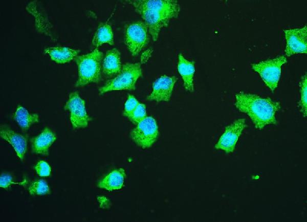
ARG59688 anti-HSPA2 antibody [4A4] ICC/IF image
Immunofluorescence: PC-3 cells were blocked with 10% goat serum and then stained with ARG59688 anti-HSPA2 antibody [4A4] (green) at 2 µg/ml dilution, overnight at 4°C. DAPI (blue) for nuclear staining.
-
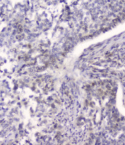
ARG59688 anti-HSPA2 antibody [4A4] IHC-P image
Immunohistochemistry: Paraffin-embedded Human lung cancer tissue. Antigen Retrieval: Heat mediation was performed in Citrate buffer (pH 6.0, epitope retrieval solution) for 20 min. The tissue section was blocked with 10% goat serum. The tissue section was then stained with ARG59688 anti-HSPA2 antibody [4A4] at 2 µg/ml, overnight at 4°C.
-
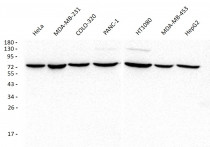
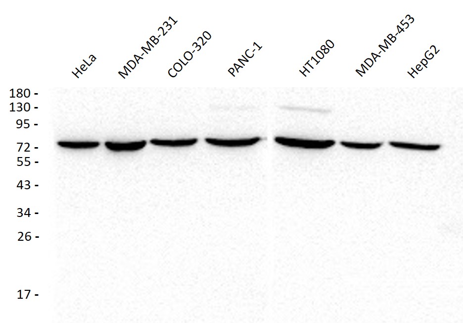
ARG59688 anti-HSPA2 antibody [4A4] WB image
Western blot: 50 µg of samples under reducing conditions. HeLa, MDA-MB-231, COLO-320, PANC-1, HT1080, MDA-MB-453 and HepG2 whole cell lysates stained with ARG59688 anti-HSPA2 antibody [4A4] at 0.5 µg/ml, overnight at 4°C.
-

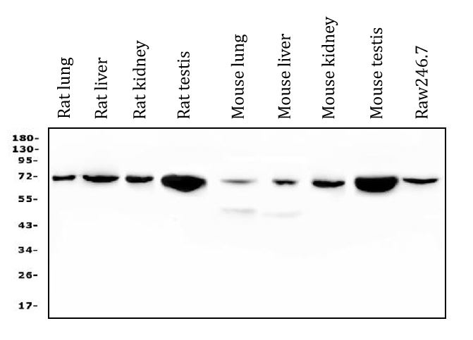
ARG59688 anti-HSPA2 antibody [4A4] WB image
Western blot: 50 µg of sample under reducing conditions. Rat lung, Rat liver, Rat kidney, Rat testis, Mouse lung, Mouse liver, Mouse kidney, Mouse testis and Raw246.7 whole cell lysates stained with ARG59688 anti-HSPA2 antibody [4A4] at 0.5 µg/ml dilution, overnight at 4°C.
-
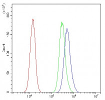
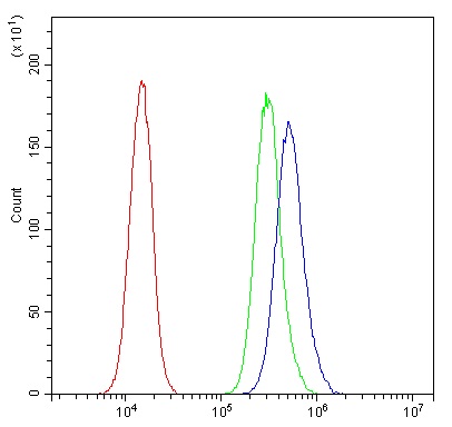
ARG59688 anti-HSPA2 antibody [4A4] FACS image
Flow Cytometry: PC-3 cells were blocked with 10% normal goat serum and then stained with ARG59688 anti-HSPA2 antibody [4A4] (blue) at 1 µg/10^6 cells for 30 min at 20°C, followed by incubation with DyLight®488 labelled secondary antibody. Isotype control antibody (green) was rabbit IgG (1 µg/10^6 cells) used under the same conditions. Unlabelled sample (red) was also used as a control.