ARG42360
anti-HLA E antibody [3D12] (PE)
anti-HLA E antibody [3D12] (PE) for Flow cytometry and Human
Overview
| Product Description | PE-conjugated Mouse Monoclonal antibody [3D12] recognizes HLA E |
|---|---|
| Tested Reactivity | Hu |
| Tested Application | FACS |
| Specificity | The mouse monoclonal antibody 3D12 (also known as 3D12HLA-E) recognizes native extracellular part of HLA-E, an ubiquitously expressed non-classical MHC class I molecule, as well as free HLA-E. |
| Host | Mouse |
| Clonality | Monoclonal |
| Clone | 3D12 |
| Isotype | IgG1 |
| Target Name | HLA E |
| Antigen Species | Human |
| Immunogen | Human HLA E. |
| Conjugation | PE |
| Alternate Names | MHC class I antigen E; QA1; EA2.1; HLA-6.2; EA1.2; MHC; HLA class I histocompatibility antigen, alpha chain E |
Application Instructions
| Application Suggestion |
|
||||
|---|---|---|---|---|---|
| Application Note | * The dilutions indicate recommended starting dilutions and the optimal dilutions or concentrations should be determined by the scientist. |
Properties
| Form | Liquid |
|---|---|
| Purification | Purified |
| Buffer | PBS and 15 mM Sodium azide. |
| Preservative | 15 mM Sodium azide |
| Concentration | 0.1 mg/ml |
| Storage Instruction | Aliquot and store in the dark at 2-8°C. Keep protected from prolonged exposure to light. Avoid repeated freeze/thaw cycles. Suggest spin the vial prior to opening. The antibody solution should be gently mixed before use. |
| Note | For laboratory research only, not for drug, diagnostic or other use. |
Bioinformation
| Database Links |
Swiss-port # P13747 Human HLA class I histocompatibility antigen, alpha chain E |
|---|---|
| Gene Symbol | HLA-E |
| Gene Full Name | major histocompatibility complex, class I, E |
| Background | HLA-E belongs to the HLA class I heavy chain paralogues. This class I molecule is a heterodimer consisting of a heavy chain and a light chain (beta-2 microglobulin). The heavy chain is anchored in the membrane. HLA-E binds a restricted subset of peptides derived from the leader peptides of other class I molecules. The heavy chain is approximately 45 kDa and its gene contains 8 exons. Exon one encodes the leader peptide, exons 2 and 3 encode the alpha1 and alpha2 domains, which both bind the peptide, exon 4 encodes the alpha3 domain, exon 5 encodes the transmembrane region, and exons 6 and 7 encode the cytoplasmic tail. [provided by RefSeq, Jul 2008] |
| Function | Non-classical major histocompatibility class Ib molecule involved in immune self-nonself discrimination. In complex with B2M/beta-2-microglobulin binds nonamer self-peptides derived from the signal sequence of classical MHC class Ia molecules (VL9 peptides) (PubMed:9754572, PubMed:18083576, PubMed:18339401). Peptide-bound HLA-E-B2M heterotrimeric complex primarily functions as a ligand for natural killer (NK) cell inhibitory receptor KLRD1-KLRC1, enabling NK cells to monitor the expression of other MHC class I molecules in healthy cells and to tolerate self (PubMed:9754572, PubMed:9486650, PubMed:17179229, PubMed:18083576). Upon cellular stress, preferentially binds signal sequence-derived peptides from stress-induced chaperones and is no longer recognized by NK cell inhibitory receptor KLRD1-KLRC1, resulting in impaired protection from NK cells (PubMed:12461076). Binds signal sequence-derived peptides from non-classical MHC class Ib HLA-G molecules and acts as a ligand for NK cell activating receptor KLRD1-KLRC2, likely playing a role in the generation and effector functions of adaptive NK cells and in maternal-fetal tolerance during pregnancy (PubMed:9754572, PubMed:30134159). Besides self-peptides, can also bind and present pathogen-derived peptides conformationally similar to VL9 peptides to alpha-beta T cell receptor (TCR) on unconventional CD8+ cytotoxic T cells, ultimately triggering antimicrobial immune response (PubMed:16474394, PubMed:30087334). (Microbial infection) Viruses like human cytomegalovirus have evolved an escape mechanism whereby virus-induced down-regulation of host MHC class I molecules is coupled to the binding of viral peptides to HLA-E, restoring HLA-E expression and inducing HLA-E-dependent NK cell immune tolerance to infected cells. [UniProt] |
| Cellular Localization | Membrane; Single-pass type I membrane protein. [UniProt] |
| Calculated MW | 40 kDa |
Images (2) Click the Picture to Zoom In
-

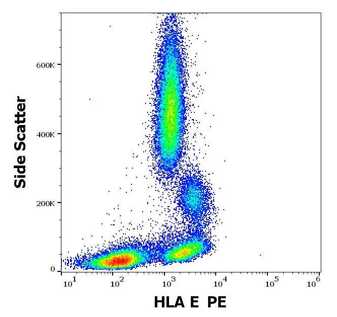
ARG42360 anti-HLA E antibody [3D12] (PE) FACS image
Flow Cytometry: Human peripheral blood stained with ARG42360 anti-HLA E antibody [3D12] (PE) at 2 µg/ml dilution.
-
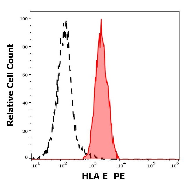
ARG42360 anti-HLA E antibody [3D12] (PE) FACS image
Flow Cytometry: Separation of Human lymphocytes (red-filled) from blood debris (black-dashed). Human peripheral whole blood stained with ARG42360 anti-HLA E antibody [3D12] (PE) at 2 µg/ml dilution.