ARG63009
anti-HLA DR antibody [MEM-12] (FITC)
anti-HLA DR antibody [MEM-12] (FITC) for Flow cytometry and Human
Immune System antibody
Overview
| Product Description | FITC-conjugated Mouse Monoclonal antibody [MEM-12] recognizes HLA DR |
|---|---|
| Tested Reactivity | Hu |
| Tested Application | FACS |
| Specificity | The clone MEM-12 recognizes common epitope on human HLA-DR which is dependent on the association of alpha and beta chains. DR is the isotype of human MHC Class II molecules expressed on antigen-presenting cells (APC; dendritic cells, B lymphocytes, monocytes, macrophages). |
| Host | Mouse |
| Clonality | Monoclonal |
| Clone | MEM-12 |
| Isotype | IgG1 |
| Target Name | HLA DR |
| Immunogen | thymocyte membrane |
| Conjugation | FITC |
| Alternate Names | MLRW; HLA class II histocompatibility antigen, DR alpha chain; HLA-DRA1; MHC class II antigen DRA |
Application Instructions
| Application Suggestion |
|
||||
|---|---|---|---|---|---|
| Application Note | * The dilutions indicate recommended starting dilutions and the optimal dilutions or concentrations should be determined by the scientist. |
Properties
| Form | Liquid |
|---|---|
| Purification Note | The purified antibody is conjugated with Fluorescein isothiocyanate (FITC) under optimum conditions. The reagent is free of unconjugated FITC and adjusted for direct use. No reconstitution is necessary. |
| Buffer | PBS, 15 mM Sodium azide and 0.2% (w/v) high-grade protease free BSA |
| Preservative | 15 mM Sodium azide |
| Stabilizer | 0.2% (w/v) high-grade protease free BSA |
| Storage Instruction | Aliquot and store in the dark at 2-8°C. Keep protected from prolonged exposure to light. Avoid repeated freeze/thaw cycles. Suggest spin the vial prior to opening. The antibody solution should be gently mixed before use. |
| Note | For laboratory research only, not for drug, diagnostic or other use. |
Bioinformation
| Database Links |
Swiss-port # P01903 Human HLA class II histocompatibility antigen, DR alpha chain |
|---|---|
| Gene Symbol | HLA-DRA |
| Gene Full Name | major histocompatibility complex, class II, DR alpha |
| Background | HLA-DR, a member of MHC class II glycoproteins, that bind intracellularly processed peptides and present them to the Th cells, is composed of 36 kDa alpha chain and 27 kDa beta chain, both anchored in the plasma membrane. Together with other MHC II molecules HLA-DR plays a central role in the immune system. |
| Function | Binds peptides derived from antigens that access the endocytic route of antigen presenting cells (APC) and presents them on the cell surface for recognition by the CD4 T-cells. The peptide binding cleft accommodates peptides of 10-30 residues. The peptides presented by MHC class II molecules are generated mostly by degradation of proteins that access the endocytic route, where they are processed by lysosomal proteases and other hydrolases. Exogenous antigens that have been endocytosed by the APC are thus readily available for presentation via MHC II molecules, and for this reason this antigen presentation pathway is usually referred to as exogenous. As membrane proteins on their way to degradation in lysosomes as part of their normal turn-over are also contained in the endosomal/lysosomal compartments, exogenous antigens must compete with those derived from endogenous components. Autophagy is also a source of endogenous peptides, autophagosomes constitutively fuse with MHC class II loading compartments. In addition to APCs, other cells of the gastrointestinal tract, such as epithelial cells, express MHC class II molecules and CD74 and act as APCs, which is an unusual trait of the GI tract. To produce a MHC class II molecule that presents an antigen, three MHC class II molecules (heterodimers of an alpha and a beta chain) associate with a CD74 trimer in the ER to form a heterononamer. Soon after the entry of this complex into the endosomal/lysosomal system where antigen processing occurs, CD74 undergoes a sequential degradation by various proteases, including CTSS and CTSL, leaving a small fragment termed CLIP (class-II-associated invariant chain peptide). The removal of CLIP is facilitated by HLA-DM via direct binding to the alpha-beta-CLIP complex so that CLIP is released. HLA-DM stabilizes MHC class II molecules until primary high affinity antigenic peptides are bound. The MHC II molecule bound to a peptide is then transported to the cell membrane surface. In B-cells, the interaction between HLA-DM and MHC class II molecules is regulated by HLA-DO. Primary dendritic cells (DCs) also to express HLA-DO. Lysosomal microenvironment has been implicated in the regulation of antigen loading into MHC II molecules, increased acidification produces increased proteolysis and efficient peptide loading. [UniProt] |
| Research Area | Immune System antibody |
| Calculated MW | 29 kDa |
| PTM | Ubiquitinated by MARCH1 or MARCH8 at Lys-244 leading to down-regulation of MHC class II. When associated with ubiquitination of the beta subunit of HLA-DR: HLA-DRB4 'Lys-254', the down-regulation of MHC class II may be highly effective. |
Images (3) Click the Picture to Zoom In
-

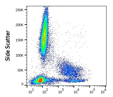
ARG63009 anti-HLA DR antibody [MEM-12] (FITC) FACS image
Flow Cytometry: Human peripheral whole blood stained with ARG63009 anti-HLA DR antibody [MEM-12] (FITC) (20 µl reagent / 100 µl of peripheral whole blood).
-

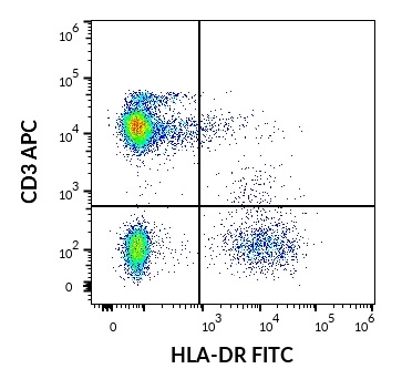
ARG63009 anti-HLA DR antibody [MEM-12] (FITC) FACS image
Flow Cytometry: Human lymphocytes stained with ARG63009 anti-HLA DR antibody [MEM-12] (FITC) (20 µl reagent / 100 µl of peripheral whole blood) and ARG54302 anti-CD3 antibody [UCHT1] (APC) (10 µl reagent / 100 µl of peripheral whole blood).
-
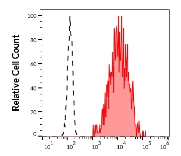
ARG63009 anti-HLA DR antibody [MEM-12] (FITC) FACS image
Flow Cytometry: Separation of human HLA-DR positive CD3 negative lymphocytes (red-filled) from neutrophil granulocytes (black-dashed). Human peripheral whole blood stained with ARG63009 anti-HLA DR antibody [MEM-12] (FITC) (20 µl reagent / 100 µl of peripheral whole blood).