ARG63006
anti-HLA DQ1 + DQ3 antibody [HL-37]
anti-HLA DQ1 + DQ3 antibody [HL-37] for Flow cytometry,Immunoprecipitation,Western blot and Human,Pig
Immune System antibody
Overview
| Product Description | Mouse Monoclonal antibody [HL-37] recognizes HLA DQ1 + DQ3 |
|---|---|
| Tested Reactivity | Hu, Pig |
| Tested Application | FACS, IP, WB |
| Specificity | The clone HL-37 reacts with polymorphic determinant on human HLA-DQ1 and HLA-DQ3 molecules (recognized epitope was found on isolated beta chain of DQ1), but does not react with HLA-DQ2. DQ is the isotype of human MHC Class II molecules expressed on antigen-presenting cells (APC; dendritic cells, B lymphocytes, monocytes, macrophages). |
| Host | Mouse |
| Clonality | Monoclonal |
| Clone | HL-37 |
| Isotype | IgG3 |
| Target Name | HLA DQ1 + DQ3 |
| Antigen Species | Human |
| Immunogen | Burkitt's lymphoma cell line Raji. |
| Conjugation | Un-conjugated |
| Alternate Names | HLA-DQB; CELIAC1; MHC class II antigen DQB1; IDDM1; HLA class II histocompatibility antigen, DQ beta 1 chain |
Application Instructions
| Application Suggestion |
|
||||||||
|---|---|---|---|---|---|---|---|---|---|
| Application Note | WB: Under non-reducing condition. * The dilutions indicate recommended starting dilutions and the optimal dilutions or concentrations should be determined by the scientist. |
Properties
| Form | Liquid |
|---|---|
| Purification | Purified from ascites by protein-A affinity chromatography. |
| Purity | > 95% (by SDS-PAGE) |
| Buffer | PBS (pH 7.4) and 15 mM Sodium azide |
| Preservative | 15 mM Sodium azide |
| Concentration | 1 mg/ml |
| Storage Instruction | For continuous use, store undiluted antibody at 2-8°C for up to a week. For long-term storage, aliquot and store at -20°C or below. Storage in frost free freezers is not recommended. Avoid repeated freeze/thaw cycles. Suggest spin the vial prior to opening. The antibody solution should be gently mixed before use. |
| Note | For laboratory research only, not for drug, diagnostic or other use. |
Bioinformation
| Database Links |
Swiss-port # P01920 Human HLA class II histocompatibility antigen, DQ beta 1 chain |
|---|---|
| Gene Symbol | HLA-DQB1 |
| Gene Full Name | major histocompatibility complex, class II, DQ beta 1 |
| Background | HLA-DQB1 belongs to the HLA class II beta chain paralogs. This class II molecule is a heterodimer consisting of an alpha (DQA) and a beta chain (DQB), both anchored in the membrane. It plays a central role in the immune system by presenting peptides derived from extracellular proteins. Class II molecules are expressed in antigen presenting cells (APC: B lymphocytes, dendritic cells, macrophages). The beta chain is approximately 26-28 kDa and it contains six exons. Exon 1 encodes the leader peptide, exons 2 and 3 encode the two extracellular domains, exon 4 encodes the transmembrane domain and exon 5 encodes the cytoplasmic tail. Within the DQ molecule both the alpha chain and the beta chain contain the polymorphisms specifying the peptide binding specificities, resulting in up to four different molecules. Typing for these polymorphisms is routinely done for bone marrow transplantation. Alternative splicing results in multiple transcript variants. [provided by RefSeq, Sep 2011] |
| Function | Binds peptides derived from antigens that access the endocytic route of antigen presenting cells (APC) and presents them on the cell surface for recognition by the CD4 T-cells. The peptide binding cleft accommodates peptides of 10-30 residues. The peptides presented by MHC class II molecules are generated mostly by degradation of proteins that access the endocytic route, where they are processed by lysosomal proteases and other hydrolases. Exogenous antigens that have been endocytosed by the APC are thus readily available for presentation via MHC II molecules, and for this reason this antigen presentation pathway is usually referred to as exogenous. As membrane proteins on their way to degradation in lysosomes as part of their normal turn-over are also contained in the endosomal/lysosomal compartments, exogenous antigens must compete with those derived from endogenous components. Autophagy is also a source of endogenous peptides, autophagosomes constitutively fuse with MHC class II loading compartments. In addition to APCs, other cells of the gastrointestinal tract, such as epithelial cells, express MHC class II molecules and CD74 and act as APCs, which is an unusual trait of the GI tract. To produce a MHC class II molecule that presents an antigen, three MHC class II molecules (heterodimers of an alpha and a beta chain) associate with a CD74 trimer in the ER to form a heterononamer. Soon after the entry of this complex into the endosomal/lysosomal system where antigen processing occurs, CD74 undergoes a sequential degradation by various proteases, including CTSS and CTSL, leaving a small fragment termed CLIP (class-II-associated invariant chain peptide). The removal of CLIP is facilitated by HLA-DM via direct binding to the alpha-beta-CLIP complex so that CLIP is released. HLA-DM stabilizes MHC class II molecules until primary high affinity antigenic peptides are bound. The MHC II molecule bound to a peptide is then transported to the cell membrane surface. In B-cells, the interaction between HLA-DM and MHC class II molecules is regulated by HLA-DO. Primary dendritic cells (DCs) also to express HLA-DO. Lysosomal microenvironment has been implicated in the regulation of antigen loading into MHC II molecules, increased acidification produces increased proteolysis and efficient peptide loading. [UniProt] |
| Research Area | Immune System antibody |
| Calculated MW | 30 kDa |
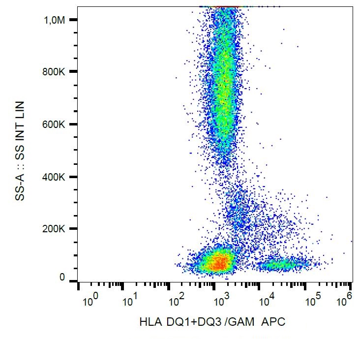
Images (1) Click the Picture to Zoom In
-

ARG63006 anti-HLA DQ1 + DQ3 antibody [HL-37] FACS image
Flow Cytometry: Human peripheral blood cells stained with ARG63006 anti-HLA DQ1 + DQ3 antibody [HL-37], followed by incubation with APC labelled Goat anti-Mouse secondary antibody.