ARG58031
anti-HINT1 antibody
anti-HINT1 antibody for Flow cytometry,ICC/IF,IHC-Formalin-fixed paraffin-embedded sections,Western blot and Human,Mouse,Rat
Overview
| Product Description | Rabbit Polyclonal antibody recognizes HINT1 |
|---|---|
| Tested Reactivity | Hu, Ms, Rat |
| Tested Application | FACS, ICC/IF, IHC-P, WB |
| Host | Rabbit |
| Clonality | Polyclonal |
| Isotype | IgG |
| Target Name | HINT1 |
| Antigen Species | Human |
| Immunogen | Synthetic peptide from Human HINT1. (HISQISVAEDDDESLLGHLMIVGKKCAADLGLNK) |
| Conjugation | Un-conjugated |
| Alternate Names | Adenosine 5'-monophosphoramidase; Protein kinase C inhibitor 1; PKCI-1; Protein kinase C-interacting protein 1; HINT; NMAN; EC 3.-.-.-; Histidine triad nucleotide-binding protein 1; PRKCNH1 |
Application Instructions
| Application Suggestion |
|
||||||||||
|---|---|---|---|---|---|---|---|---|---|---|---|
| Application Note | IHC-P: Antigen Retrieval: Boil tissue section in 10 mM Citrate buffer (pH 6.0) for 20 min, followed by cooling at RT. * The dilutions indicate recommended starting dilutions and the optimal dilutions or concentrations should be determined by the scientist. |
||||||||||
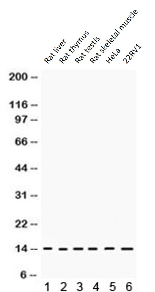
| Observed Size | ~ 14 kDa |
Properties
| Form | Liquid |
|---|---|
| Purification | Affinity purification with immunogen. |
| Buffer | PBS, 0.025% Sodium azide and 2.5% BSA. |
| Preservative | 0.025% Sodium azide |
| Stabilizer | 2.5% BSA |
| Concentration | 0.5 mg/ml |
| Storage Instruction | For continuous use, store undiluted antibody at 2-8°C for up to a week. For long-term storage, aliquot and store at -20°C or below. Storage in frost free freezers is not recommended. Avoid repeated freeze/thaw cycles. Suggest spin the vial prior to opening. The antibody solution should be gently mixed before use. |
| Note | For laboratory research only, not for drug, diagnostic or other use. |
Bioinformation
| Database Links | |
|---|---|
| Gene Symbol | HINT1 |
| Gene Full Name | histidine triad nucleotide binding protein 1 |
| Background | The protein encoded by this gene can hydrolyze substrates such as AMP-morpholidate, AMP-N-alanine methyl ester, AMP-alpha-acetyl lysine methyl ester, and AMP-NH2. The encoded protein interacts with these substrates via a histidine triad motif, which is part of the loop that binds to the substrate. This gene has been found to be a tumor suppressing gene. Several transcript variants, but only one of them protein-coding, have been found for this gene. [provided by RefSeq, Dec 2012] |
| Function | Hydrolyzes purine nucleotide phosphoramidates with a single phosphate group, including adenosine 5'monophosphoramidate (AMP-NH2), adenosine 5'monophosphomorpholidate (AMP-morpholidate) and guanosine 5'monophosphomorpholidate (GMP-morpholidate). Hydrolyzes lysyl-AMP (AMP-N-epsilon-(N-alpha-acetyl lysine methyl ester)) generated by lysine tRNA ligase, as well as Met-AMP, His-AMP and Asp-AMP, lysyl-GMP (GMP-N-epsilon-(N-alpha-acetyl lysine methyl ester)) and AMP-N-alanine methyl ester. Can also convert adenosine 5'-O-phosphorothioate and guanosine 5'-O-phosphorothioate to the corresponding nucleoside 5'-O-phosphates with concomitant release of hydrogen sulfide. In addition, functions as scaffolding protein that modulates transcriptional activation by the LEF1/TCF1-CTNNB1 complex and by the complex formed with MITF and CTNNB1. Modulates p53/TP53 levels and p53/TP53-mediated apoptosis. Modulates proteasomal degradation of target proteins by the SCF (SKP2-CUL1-F-box protein) E3 ubiquitin-protein ligase complex. [UniProt] |
| Calculated MW | 14 kDa |
Images (6) Click the Picture to Zoom In
-
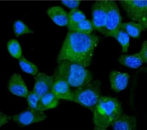
ARG58031 anti-HINT1 antibody ICC/IF image
Immunofluorescence: A431 cells stained with ARG58031 anti-HINT1 antibody (green). DAPI (blue) for nuclear staining.
-
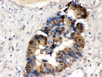
ARG58031 anti-HINT1 antibody IHC-P image
Immunohistochemistry: Formalin-fixed and paraffin-embedded Human intestinal cancer tissue stained with ARG58031 anti-HINT1 antibody. Antigen Retrieval: Boil tissue section in 10mM Citrate buffer (pH 6.0) for 20 min followed by cooling at RT.
-
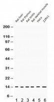
ARG58031 anti-HINT1 antibody WB image
Western blot: 1) Rat liver, 2) Rat thymus, 3) Rat testis, 4) Rat skeletal muscle, 5) HeLa, 6) 22RV1 lysates stained with ARG58031 anti-HINT1 antibody.
-
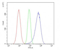
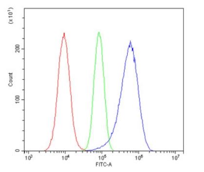
ARG58031 anti-HINT1 antibody FACS image
Flow Cytometry: A549 cells were blocked with goat sera and stained with ARG58031 anti-HINT1 antibody at 1 µg/10^6 cells (blue); Cells alone (red); Isotype control (green).
-
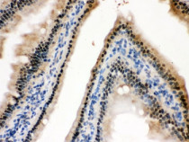
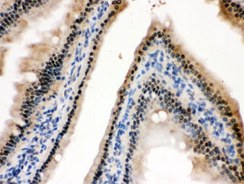
ARG58031 anti-HINT1 antibody IHC-P image
Immunohistochemistry: Formalin-fixed and paraffin-embedded Mouse intestine stained with ARG58031 anti-HINT1 antibody. Antigen Retrieval: Boil tissue section in 10mM Citrate buffer (pH 6.0) for 20 min followed by cooling at RT.
-
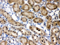
ARG58031 anti-HINT1 antibody IHC-P image
Immunohistochemistry: Formalin-fixed and paraffin-embedded Rat kidney stained with ARG58031 anti-HINT1 antibody. Antigen Retrieval: Boil tissue section in 10mM Citrate buffer (pH 6.0) for 20 min followed by cooling at RT.