ARG40688
anti-HDAC4 antibody
anti-HDAC4 antibody for Flow cytometry,ICC/IF,IHC-Formalin-fixed paraffin-embedded sections,Western blot and Human,Mouse,Rat
Overview
| Product Description | Rabbit Polyclonal antibody recognizes HDAC4 |
|---|---|
| Tested Reactivity | Hu, Ms, Rat |
| Tested Application | FACS, ICC/IF, IHC-P, WB |
| Host | Rabbit |
| Clonality | Polyclonal |
| Isotype | IgG |
| Target Name | HDAC4 |
| Antigen Species | Human |
| Immunogen | Recombinant protein corresponding to Q4-Q213 of Human HDAC4. |
| Conjugation | Un-conjugated |
| Alternate Names | HD4; BDMR; HDAC-A; HDACA; EC 3.5.1.98; HDAC-4; HA6116; Histone deacetylase 4; AHO3 |
Application Instructions
| Application Suggestion |
|
||||||||||
|---|---|---|---|---|---|---|---|---|---|---|---|
| Application Note | IHC-P: Antigen Retrieval: Heat mediation was performed in EDTA buffer (pH 8.0). * The dilutions indicate recommended starting dilutions and the optimal dilutions or concentrations should be determined by the scientist. |
Properties
| Form | Liquid |
|---|---|
| Purification | Affinity purification with immunogen. |
| Buffer | 0.2% Na2HPO4, 0.9% NaCl, 0.05% Sodium azide and 5% BSA. |
| Preservative | 0.05% Sodium azide |
| Stabilizer | 5% BSA |
| Concentration | 0.5 mg/ml |
| Storage Instruction | For continuous use, store undiluted antibody at 2-8°C for up to a week. For long-term storage, aliquot and store at -20°C or below. Storage in frost free freezers is not recommended. Avoid repeated freeze/thaw cycles. Suggest spin the vial prior to opening. The antibody solution should be gently mixed before use. |
| Note | For laboratory research only, not for drug, diagnostic or other use. |
Bioinformation
| Database Links | |
|---|---|
| Gene Symbol | HDAC4 |
| Gene Full Name | histone deacetylase 4 |
| Background | Histones play a critical role in transcriptional regulation, cell cycle progression, and developmental events. Histone acetylation/deacetylation alters chromosome structure and affects transcription factor access to DNA. The protein encoded by this gene belongs to class II of the histone deacetylase/acuc/apha family. It possesses histone deacetylase activity and represses transcription when tethered to a promoter. This protein does not bind DNA directly, but through transcription factors MEF2C and MEF2D. It seems to interact in a multiprotein complex with RbAp48 and HDAC3. [provided by RefSeq, Jul 2008] |
| Function | Responsible for the deacetylation of lysine residues on the N-terminal part of the core histones (H2A, H2B, H3 and H4). Histone deacetylation gives a tag for epigenetic repression and plays an important role in transcriptional regulation, cell cycle progression and developmental events. Histone deacetylases act via the formation of large multiprotein complexes. Involved in muscle maturation via its interaction with the myocyte enhancer factors such as MEF2A, MEF2C and MEF2D. Involved in the MTA1-mediated epigenetic regulation of ESR1 expression in breast cancer. [UniProt] |
| Cellular Localization | Nucleus. Cytoplasm. Note=Shuttles between the nucleus and the cytoplasm. Upon muscle cells differentiation, it accumulates in the nuclei of myotubes, suggesting a positive role of nuclear HDAC4 in muscle differentiation. The export to cytoplasm depends on the interaction with a 14-3-3 chaperone protein and is due to its phosphorylation at Ser-246, Ser-467 and Ser-632 by CaMK4 and SIK1. The nuclear localization probably depends on sumoylation. [UniProt] |
| Calculated MW | 119 kDa |
| PTM | Phosphorylated by CaMK4 at Ser-246, Ser-467 and Ser-632. Phosphorylation at other residues by CaMK2D is required for the interaction with 14-3-3. Phosphorylation at Ser-350, within the PxLPxI/L motif, impairs the binding of ANKRA2 but generates a high-affinity docking site for 14-3-3. Sumoylation on Lys-559 is promoted by the E3 SUMO-protein ligase RANBP2, and prevented by phosphorylation by CaMK4. [UniProt] |
Images (7) Click the Picture to Zoom In
-

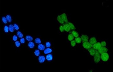
ARG40688 anti-HDAC4 antibody ICC/IF image
Immunofluorescence: MCF-7 cells were blocked with 10% goat serum and then stained with ARG40688 anti-HDAC4 antibody (green) at 5 µg/ml dilution, overnight at 4°C. DAPI (blue) for nuclear staining.
-
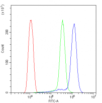
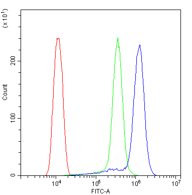
ARG40688 anti-HDAC4 antibody FACS image
Flow Cytometry: THP-1 cells were blocked with 10% normal goat serum and stained with ARG40688 anti-HDAC4 antibody (blue) at 1 µg/10^6 cells for 30 min at 20°C, followed by incubation with DyLight®488 labelled secondary antibody. Isotype control antibody (green) was rabbit IgG (1 µg/10^6 cells) used under the same conditions. Unlabelled sample (red) was also used as a control.
-
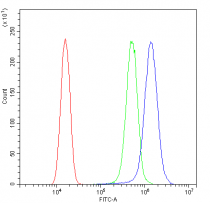
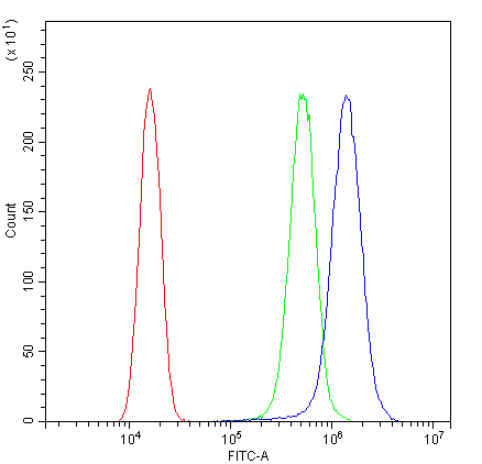
ARG40688 anti-HDAC4 antibody FACS image
Flow Cytometry: SiHa cells were blocked with 10% normal goat serum and stained with ARG40688 anti-HDAC4 antibody (blue) at 1 µg/10^6 cells for 30 min at 20°C, followed by incubation with DyLight®488 labelled secondary antibody. Isotype control antibody (green) was rabbit IgG (1 µg/10^6 cells) used under the same conditions. Unlabelled sample (red) was also used as a control.
-
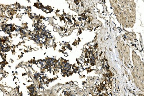
ARG40688 anti-HDAC4 antibody IHC-P image
Immunohistochemistry: Paraffin-embedded Human bladder cancer tissue. Antigen Retrieval: Heat mediation was performed in EDTA buffer (pH 8.0). The tissue section was blocked with 10% goat serum. The tissue section was then stained with ARG40688 anti-HDAC4 antibody at 2 µg/ml dilution, overnight at 4°C.
-
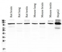
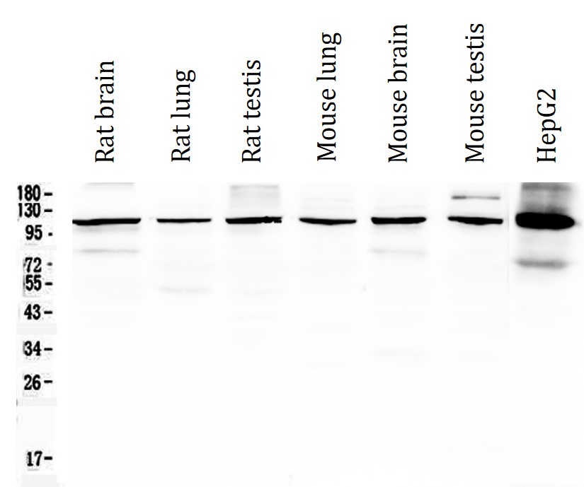
ARG40688 anti-HDAC4 antibody WB image
Western blot: 50 µg of samples under reducing conditions. Rat brain, Rat lung, Rat testis, Mouse lung, Mouse brain, Mouse testis and HepG2 whole cell lysates stained with ARG40688 anti-HDAC4 antibody at 0.5 µg/ml, overnight at 4°C.
-
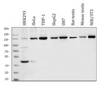
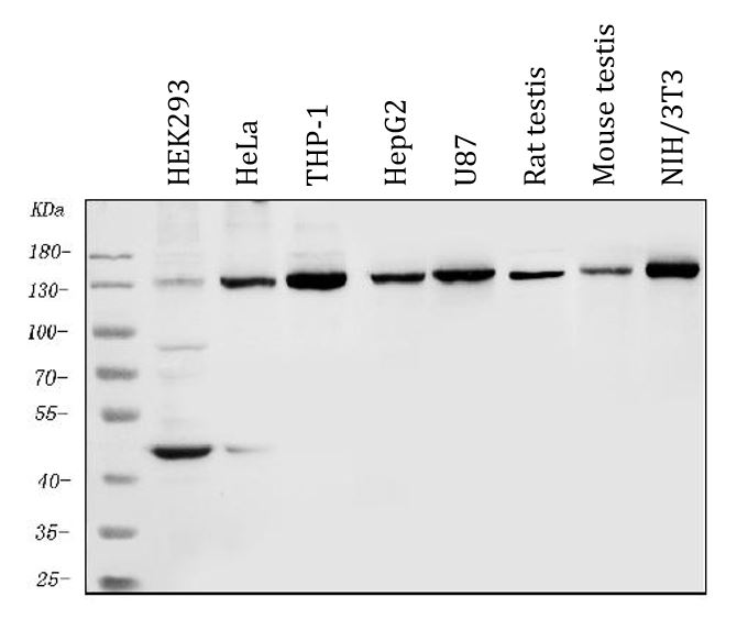
ARG40688 anti-HDAC4 antibody WB image
Western blot: 50 µg of sample under reducing conditions. HEK293, HeLa, THP-1, HepG2, U87, Rat testis, Mouse testis and NIH/3T3 whole cell lysates stained with ARG40688 anti-HDAC4 antibody at 0.5 µg/ml dilution, overnight at 4°C.
-

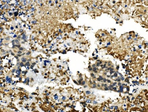
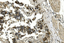
ARG40688 anti-HDAC4 antibody IHC-P image
Immunohistochemistry: Paraffin-embedded Human pancreatic cancer tissue. Antigen Retrieval: Heat mediation was performed in EDTA buffer (pH 8.0). The tissue section was blocked with 10% goat serum. The tissue section was then stained with ARG40688 anti-HDAC4 antibody at 2 µg/ml dilution, overnight at 4°C.