ARG55389
anti-HDAC1 antibody
anti-HDAC1 antibody for ICC/IF,IHC-Formalin-fixed paraffin-embedded sections,Immunoprecipitation,Western blot and Human,Mouse,Rat
Cancer antibody; Cell Biology and Cellular Response antibody; Cell Death antibody; Gene Regulation antibody

Overview
| Product Description | Rabbit Polyclonal antibody recognizes HDAC1 |
|---|---|
| Tested Reactivity | Hu, Ms, Rat |
| Tested Application | ICC/IF, IHC-P, IP, WB |
| Host | Rabbit |
| Clonality | Polyclonal |
| Isotype | IgG |
| Target Name | HDAC1 |
| Antigen Species | Human |
| Immunogen | Recombinant protein of Human HDAC1 (NP_004955.2). |
| Conjugation | Un-conjugated |
| Alternate Names | EC 3.5.1.98; HD1; RPD3L1; Histone deacetylase 1; GON-10; RPD3 |
Application Instructions
| Application Suggestion |
|
||||||||||
|---|---|---|---|---|---|---|---|---|---|---|---|
| Application Note | * The dilutions indicate recommended starting dilutions and the optimal dilutions or concentrations should be determined by the scientist. |
Properties
| Form | Liquid |
|---|---|
| Purification | Affinity purification with immunogen. |
| Buffer | PBS (pH 7.3), 0.02% Sodium azide and 50% Glycerol |
| Preservative | 0.02% Sodium azide |
| Stabilizer | 50% Glycerol |
| Storage Instruction | For continuous use, store undiluted antibody at 2-8°C for up to a week. For long-term storage, aliquot and store at -20°C. Storage in frost free freezers is not recommended. Avoid repeated freeze/thaw cycles. Suggest spin the vial prior to opening. The antibody solution should be gently mixed before use. |
| Note | For laboratory research only, not for drug, diagnostic or other use. |
Bioinformation
| Database Links | |
|---|---|
| Gene Symbol | HDAC1 |
| Gene Full Name | histone deacetylase 1 |
| Background | Histone acetylation and deacetylation, catalyzed by multisubunit complexes, play a key role in the regulation of eukaryotic gene expression. The protein encoded by this gene belongs to the histone deacetylase/acuc/apha family and is a component of the histone deacetylase complex. It also interacts with retinoblastoma tumor-suppressor protein and this complex is a key element in the control of cell proliferation and differentiation. Together with metastasis-associated protein-2, it deacetylates p53 and modulates its effect on cell growth and apoptosis. [provided by RefSeq, Jul 2008] |
| Function | Responsible for the deacetylation of lysine residues on the N-terminal part of the core histones (H2A, H2B, H3 and H4). Histone deacetylation gives a tag for epigenetic repression and plays an important role in transcriptional regulation, cell cycle progression and developmental events. Histone deacetylases act via the formation of large multiprotein complexes. Deacetylates SP proteins, SP1 and SP3, and regulates their function. Component of the BRG1-RB1-HDAC1 complex, which negatively regulates the CREST-mediated transcription in resting neurons. Upon calcium stimulation, HDAC1 is released from the complex and CREBBP is recruited, which facilitates transcriptional activation. Deacetylates TSHZ3 and regulates its transcriptional repressor activity. Deacetylates 'Lys-310' in RELA and thereby inhibits the transcriptional activity of NF-kappa-B. Deacetylates NR1D2 and abrogates the effect of KAT5-mediated relieving of NR1D2 transcription repression activity. Component of a RCOR/GFI/KDM1A/HDAC complex that suppresses, via histone deacetylase (HDAC) recruitment, a number of genes implicated in multilineage blood cell development. Involved in CIART-mediated transcriptional repression of the circadian transcriptional activator: CLOCK-ARNTL/BMAL1 heterodimer. Required for the transcriptional repression of circadian target genes, such as PER1, mediated by the large PER complex or CRY1 through histone deacetylation. [UniProt] |
| Research Area | Cancer antibody; Cell Biology and Cellular Response antibody; Cell Death antibody; Gene Regulation antibody |
| Calculated MW | 55 kDa |
| PTM | Sumoylated on Lys-444 and Lys-476; which promotes enzymatic activity. Desumoylated by SENP1. Phosphorylation on Ser-421 and Ser-423 promotes enzymatic activity and interactions with NuRD and SIN3 complexes. Phosphorylated by CDK5. Ubiquitinated by CHFR, leading to its degradation by the proteasome. Ubiquitinated by KCTD11, leading to proteasomal degradation. |
Images (4) Click the Picture to Zoom In
-

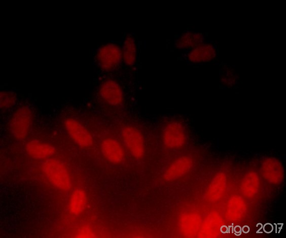
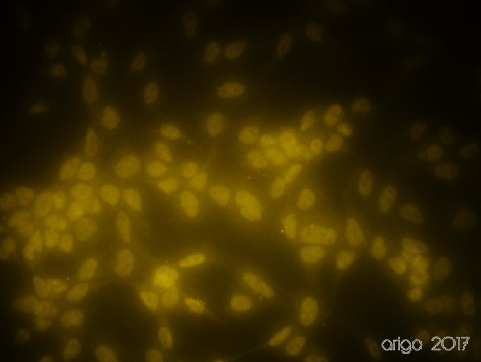
ARG55389 anti-HDAC1 antibody ICC/IF image
Immunofluorescence: 100% Methanol fixed (RT, 10 min) HeLa cells stained with ARG55389 anti-HDAC1 antibody (red) at 1:50 dilution.
Secondary antibody: ARG21917 Goat anti-Rabbit IgG antibody (TRITC)
-
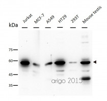
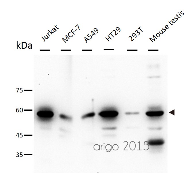
ARG55389 anti-HDAC1 antibody WB image
Western blot: 30 µg of Jurkat, MCF-7, A549, HT29, 293T and Mouse testis lysates stained with ARG55389 anti-HDAC1 antibody at 1:500 dilution.
-
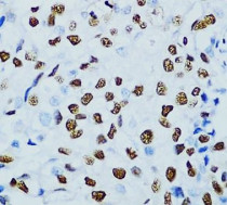
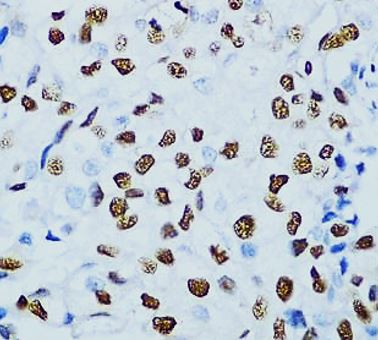
ARG55389 anti-HDAC1 antibody IHC-P image
Immunohistochemistry: Paraffin-embedded Human prostate cancer tissue stained with ARG55389 anti-HDAC1 antibody at 1:100 dilution.
-
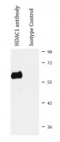
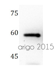
ARG55389 anti-HDAC1 antibody IP image
Immunoprecipitation: 200 µg extracts of HT-29 cells were immunoprecipitated and stained with ARG55389 anti-HDAC1 antibody at 1:1000 dilution.

Customer's Feedback
Excellent

Review for anti-HDAC1 antibody
Application:IF/ICC
Sample:HeLa
Fixation Buffer:100% Methanol
Fixation Time:10 min
Fixation Temperature:RT ºC
Permeabilization Buffer:0.1% Triton X-100
Primary Antibody Dilution Factor:1:50
Primary Antibody Incubation Time:overnight
Primary Antibody Incubation Temperature:4 ºC
Conjugation of Secondary Antibody:TRITC

Excellent
Review for anti-HDAC1 antibody
Application:IF/ICC
Sample:HeLa
Fixation Buffer:100% Methanol
Fixation Time:10 min
Fixation Temperature:RT ºC
Permeabilization Buffer:0.1% Triton X-100
Primary Antibody Dilution Factor:1:50
Primary Antibody Incubation Time:overnight
Primary Antibody Incubation Temperature:4 ºC
Conjugation of Secondary Antibody:TRITC
Excellent
Review for anti-HDAC1 antibody
Application:WB
Sample:HeLa cell lysate
Sample Loading Amount:30 µg
Primary Antibody Dilution Factor:1:500
Primary Antibody Incubation Time:overnight
Primary Antibody Incubation Temperature:4 ºC


