ARG57096
anti-HAX1 antibody [3C5]
anti-HAX1 antibody [3C5] for ICC/IF,Western blot and Human

Overview
| Product Description | Mouse Monoclonal antibody [3C5] recognizes HAX1 |
|---|---|
| Tested Reactivity | Hu |
| Tested Application | ICC/IF, WB |
| Host | Mouse |
| Clonality | Monoclonal |
| Clone | 3C5 |
| Isotype | IgG2b, kappa |
| Target Name | HAX1 |
| Antigen Species | Human |
| Immunogen | Recombinant fragment around aa. 1-279 of Human HAX1 |
| Conjugation | Un-conjugated |
| Alternate Names | HCLS1-associated protein X-1; HS1BP1; HAX-1; HS1-binding protein 1; HS1-associating protein X-1; HSP1BP-1; SCN3; HCLSBP1 |
Application Instructions
| Application Suggestion |
|
||||||
|---|---|---|---|---|---|---|---|
| Application Note | * The dilutions indicate recommended starting dilutions and the optimal dilutions or concentrations should be determined by the scientist. |
Properties
| Form | Liquid |
|---|---|
| Purification | Purification with Protein G. |
| Buffer | PBS (pH 7.4), 0.02% Sodium azide and 10% Glycerol. |
| Preservative | 0.02% Sodium azide |
| Stabilizer | 10% Glycerol |
| Concentration | 1 mg/ml |
| Storage Instruction | For continuous use, store undiluted antibody at 2-8°C for up to a week. For long-term storage, aliquot and store at -20°C. Storage in frost free freezers is not recommended. Avoid repeated freeze/thaw cycles. Suggest spin the vial prior to opening. The antibody solution should be gently mixed before use. |
| Note | For laboratory research only, not for drug, diagnostic or other use. |
Bioinformation
| Database Links | |
|---|---|
| Gene Symbol | HAX1 |
| Gene Full Name | HCLS1 associated protein X-1 |
| Background | The protein encoded by this gene is known to associate with hematopoietic cell-specific Lyn substrate 1, a substrate of Src family tyrosine kinases. It also interacts with the product of the polycystic kidney disease 2 gene, mutations in which are associated with autosomal-dominant polycystic kidney disease, and with the F-actin-binding protein, cortactin. It was earlier thought that this gene product is mainly localized in the mitochondria, however, recent studies indicate it to be localized in the cell body. Mutations in this gene result in autosomal recessive severe congenital neutropenia, also known as Kostmann disease. Two transcript variants encoding different isoforms have been found for this gene. [provided by RefSeq, Jul 2008] |
| Function | Promotes cell survival. Potentiates GNA13-mediated cell migration. Involved in the clathrin-mediated endocytosis pathway. May be involved in internalization of ABC transporters such as ABCB11. May inhibit CASP9 and CASP3. May regulate intracellular calcium pools. [UniProt] |
| Calculated MW | 32 kDa |
| PTM | Proteolytically cleaved by caspase-3 during apoptosis. |
Images (3) Click the Picture to Zoom In
-
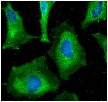
ARG57096 anti-HAX1 antibody [3C5] ICC/IF image
Immunofluorescence: HeLa cells line stained with ARG57096 anti-HAX1 antibody [3C5] at 1:100 (Green).
DAPI (Blue) for nucleus staining.
-
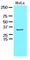
ARG57096 anti-HAX1 antibody [3C5] WB image
Western blot: 30 µg of HeLa cell lysate stained with ARG57096 anti-HAX1 antibody [3C5] at 1:1000.
-
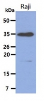
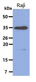
ARG57096 anti-HAX1 antibody [3C5] WB image
Western blot: 40 µg of Raji cell lysate stained with ARG57096 anti-HAX1 antibody [3C5] at 1:1000.

Customer's Feedback
Excellent
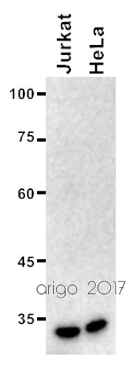
Review for anti-HAX1 antibody [3C5]
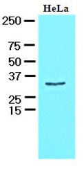
Application:WB
Sample:Jurkat and HeLa
Sample Loading Amount:20 µg
Primary Antibody Dilution Factor:1:2000
Primary Antibody Incubation Time:overnight
Primary Antibody Incubation Temperature:4 ºC


