ARG52316
anti-GluR1 Subunit phospho (Ser845) antibody
anti-GluR1 Subunit phospho (Ser845) antibody for IHC-Frozen sections,Western blot and Rat
Neuroscience antibody; Postsynaptic Receptor antibody
Overview
| Product Description | Rabbit Polyclonal antibody recognizes GluR1 Subunit phospho (Ser845) |
|---|---|
| Tested Reactivity | Rat |
| Predict Reactivity | Hu, Ms, NHuPrm |
| Tested Application | IHC-Fr, WB |
| Host | Rabbit |
| Clonality | Polyclonal |
| Isotype | IgG |
| Target Name | GluR1 Subunit |
| Antigen Species | Rat |
| Immunogen | Synthetic phospho-peptide corresponding to amino acid residues surrounding Ser845 conjugated to KLH |
| Conjugation | Un-conjugated |
| Alternate Names | GLUH1; GluA1; GluR-1; Glutamate receptor ionotropic, AMPA 1; GluR-K1; GLUR1; HBGR1; AMPA-selective glutamate receptor 1; GluR-A; GLURA; Glutamate receptor 1 |
Application Instructions
| Application Suggestion |
|
||||||
|---|---|---|---|---|---|---|---|
| Application Note | Specific for the ~100k GluR1 protein phosphorylated at Ser845 in Western blots of Rat brain extracts. Immunolabeling is blocked by the phosphopeptide used as antigen but not by the corresponding dephosphopeptide. Immunolabeling is s completely eliminated by treatment with λ-Ptase. * The dilutions indicate recommended starting dilutions and the optimal dilutions or concentrations should be determined by the scientist. |
Properties
| Form | Liquid |
|---|---|
| Purification | Affinity Purified |
| Buffer | 10 mM HEPES (pH 7.5), 150 mM NaCl, 0.1 mg/ml BSA and 50% Glycerol |
| Stabilizer | 0.1 mg/ml BSA, 50% Glycerol |
| Storage Instruction | For continuous use, store undiluted antibody at 2-8°C for up to a week. For long-term storage, aliquot and store at -20°C. Storage in frost free freezers is not recommended. Avoid repeated freeze/thaw cycles. Suggest spin the vial prior to opening. The antibody solution should be gently mixed before use. |
| Note | For laboratory research only, not for drug, diagnostic or other use. |
Bioinformation
| Database Links | |
|---|---|
| Gene Symbol | GRIA1 |
| Gene Full Name | glutamate receptor, ionotropic, AMPA 1 |
| Background | The ion channels activated by glutamate are typically divided into two classes. Those that are sensitive to N-methyl-D-aspartate (NMDA) are designated NMDA receptors (NMDAR) while those activated by α-amino-3-hydroxy-5-methyl-4-isoxalone propionic acid (AMPA) are known as AMPA receptors (AMPAR). The AMPAR are comprised of four distinct glutamate receptor subunits designated (GluR1-4) and they play key roles in virtually all excitatory neurotransmission in the brain (Keinänen et al., 1990; Hollmann and Heinemann, 1994). The GluR1 subunit is widely expressed throughout the nervous system. Phosphorylation of Ser845 on GluR1 is thought to be mediated by PKA and phosphorylation of this site increases the conductance of the AMPAR (Roche et al., 1996; Banke et al., 2000). In addition, phosphorylation of this site has been linked to synaptic plasticity as well as learning and memory (Lee at al., 2003; Esteban at al., 2003). |
| Highlight | Related Antibody Duos and Panels: ARG30132 Phospho GluR1 Antibody Panel Related products: GluR1 antibodies; GluR1 Duos / Panels; Anti-Rabbit IgG secondary antibodies; |
| Research Area | Neuroscience antibody; Postsynaptic Receptor antibody |
| Calculated MW | 102 kDa |
| PTM | Palmitoylated. Depalmitoylated upon glutamate stimulation. Cys-603 palmitoylation leads to Golgi retention and decreased cell surface expression. In contrast, Cys-829 palmitoylation does not affect cell surface expression but regulates stimulation-dependent endocytosis (By similarity). Phosphorylated at Ser-645. Phosphorylated at Ser-710 by PKC. Phosphorylated at Ser-849 by PKC, PKA and CAMK2. Phosphorylated at Ser-863 by PKC, PKA and PRKG2. |
Images (1) Click the Picture to Zoom In
-
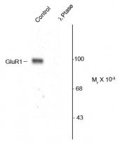
ARG52316 anti-GluR1 Subunit phospho (Ser845) antibody WB image
Western blot: Rat hippocampal lysate showing specific immunolabeling of the ~100k GluR1 protein phosphorylated at Ser845 stained with ARG52316 anti-GluR1 Subunit phospho (Ser845) antibody. The phosphospecificity of this labeling is shown in the second lane (lambda-phosphatase: λ-Ptase). The blot is identical to the control except that the second lane (lambda-phosphatase: λ-Ptase). The blot is identical to the control except that it was incubated in λ-Ptase (1200 units for 30 min) before being exposed to the primary antibody. The immunolabeling is completely eliminated by treatment with λ-Ptase.


