ARG66860
anti-GSK3 beta phospho (Ser9) antibody
anti-GSK3 beta phospho (Ser9) antibody for IHC-Formalin-fixed paraffin-embedded sections,Western blot and Human,Mouse,Rat
Overview
| Product Description | Rabbit Polyclonal antibody recognizes GSK3 beta phospho (Ser9) |
|---|---|
| Tested Reactivity | Hu, Ms, Rat |
| Tested Application | IHC-P, WB |
| Specificity | This antibody detects endogenous levels of GSK3 beta protein only when phosphorylated at Ser9. |
| Host | Rabbit |
| Clonality | Polyclonal |
| Isotype | IgG |
| Target Name | GSK3 beta |
| Antigen Species | Human |
| Immunogen | Synthetic peptide around the phosphorylated Ser9 (aa. 1-50) of Human GSK3 beta. |
| Conjugation | Un-conjugated |
| Alternate Names | EC 2.7.11.26; EC 2.7.11.1; GSK-3 beta; Glycogen synthase kinase-3 beta; Serine/threonine-protein kinase GSK3B |
Application Instructions
| Application Suggestion |
|
||||||
|---|---|---|---|---|---|---|---|
| Application Note | IHC-P: Antigen Retrieval: Boil tissue section in Sodium citrate buffer (pH 6.0) for 20 min. * The dilutions indicate recommended starting dilutions and the optimal dilutions or concentrations should be determined by the scientist. |
||||||
| Positive Control | 293 and HeLa | ||||||
| Observed Size | 42 or 47 kDa |
Properties
| Form | Liquid |
|---|---|
| Purification | Affinity purification with immunogen. |
| Buffer | PBS, 0.02% Sodium azide, 50% Glycerol and 0.5% BSA. |
| Preservative | 0.02% Sodium azide |
| Stabilizer | 50% Glycerol and 0.5% BSA |
| Concentration | 1 mg/ml |
| Storage Instruction | For continuous use, store undiluted antibody at 2-8°C for up to a week. For long-term storage, aliquot and store at -20°C. Storage in frost free freezers is not recommended. Avoid repeated freeze/thaw cycles. Suggest spin the vial prior to opening. The antibody solution should be gently mixed before use. |
| Note | For laboratory research only, not for drug, diagnostic or other use. |
Bioinformation
| Database Links | |
|---|---|
| Gene Symbol | GSK3B |
| Gene Full Name | glycogen synthase kinase 3 beta |
| Background | The protein encoded by this gene is a serine-threonine kinase belonging to the glycogen synthase kinase subfamily. It is a negative regulator of glucose homeostasis and is involved in energy metabolism, inflammation, ER-stress, mitochondrial dysfunction, and apoptotic pathways. Defects in this gene have been associated with Parkinson disease and Alzheimer disease. [provided by RefSeq, Aug 2017] |
| Function | Constitutively active protein kinase that acts as a negative regulator in the hormonal control of glucose homeostasis, Wnt signaling and regulation of transcription factors and microtubules, by phosphorylating and inactivating glycogen synthase (GYS1 or GYS2), EIF2B, CTNNB1/beta-catenin, APC, AXIN1, DPYSL2/CRMP2, JUN, NFATC1/NFATC, MAPT/TAU and MACF1. Requires primed phosphorylation of the majority of its substrates. In skeletal muscle, contributes to insulin regulation of glycogen synthesis by phosphorylating and inhibiting GYS1 activity and hence glycogen synthesis. May also mediate the development of insulin resistance by regulating activation of transcription factors. Regulates protein synthesis by controlling the activity of initiation factor 2B (EIF2BE/EIF2B5) in the same manner as glycogen synthase. In Wnt signaling, GSK3B forms a multimeric complex with APC, AXIN1 and CTNNB1/beta-catenin and phosphorylates the N-terminus of CTNNB1 leading to its degradation mediated by ubiquitin/proteasomes. Phosphorylates JUN at sites proximal to its DNA-binding domain, thereby reducing its affinity for DNA. Phosphorylates NFATC1/NFATC on conserved serine residues promoting NFATC1/NFATC nuclear export, shutting off NFATC1/NFATC gene regulation, and thereby opposing the action of calcineurin. Phosphorylates MAPT/TAU on 'Thr-548', decreasing significantly MAPT/TAU ability to bind and stabilize microtubules. MAPT/TAU is the principal component of neurofibrillary tangles in Alzheimer disease. Plays an important role in ERBB2-dependent stabilization of microtubules at the cell cortex. Phosphorylates MACF1, inhibiting its binding to microtubules which is critical for its role in bulge stem cell migration and skin wound repair. Probably regulates NF-kappa-B (NFKB1) at the transcriptional level and is required for the NF-kappa-B-mediated anti-apoptotic response to TNF-alpha (TNF/TNFA). Negatively regulates replication in pancreatic beta-cells, resulting in apoptosis, loss of beta-cells and diabetes. Through phosphorylation of the anti-apoptotic protein MCL1, may control cell apoptosis in response to growth factors deprivation. Phosphorylates MUC1 in breast cancer cells, decreasing the interaction of MUC1 with CTNNB1/beta-catenin. Is necessary for the establishment of neuronal polarity and axon outgrowth. Phosphorylates MARK2, leading to inhibit its activity. Phosphorylates SIK1 at 'Thr-182', leading to sustain its activity. Phosphorylates ZC3HAV1 which enhances its antiviral activity. Phosphorylates SNAI1, leading to its BTRC-triggered ubiquitination and proteasomal degradation. Phosphorylates SFPQ at 'Thr-687' upon T-cell activation. Phosphorylates NR1D1 st 'Ser-55' and 'Ser-59' and stabilizes it by protecting it from proteasomal degradation. Regulates the circadian clock via phosphorylation of the major clock components including ARNTL/BMAL1, CLOCK and PER2 (PubMed:19946213, PubMed:28903391). Phosphorylates CLOCK AT 'Ser-427' and targets it for proteasomal degradation (PubMed:19946213). Phosphorylates ARNTL/BMAL1 at 'Ser-17' and 'Ser-21' and primes it for ubiquitination and proteasomal degradation (PubMed:28903391). Phosphorylates OGT at 'Ser-3' or 'Ser-4' which positively regulates its activity. Phosphorylates MYCN in neuroblastoma cells which may promote its degradation (PubMed:24391509). Regulates the circadian rhythmicity of hippocampal long-term potentiation and ARNTL/BMLA1 and PER2 expression (By similarity). Acts as a regulator of autophagy by mediating phosphorylation of KAT5/TIP60 under starvation conditions, leading to activate KAT5/TIP60 acetyltransferase activity and promote acetylation of key autophagy regulators, such as ULK1 and RUBCNL/Pacer (PubMed:30704899). Negatively regulates extrinsic apoptotic signaling pathway via death domain receptors. Promotes the formation of an anti-apoptotic complex, made of DDX3X, BRIC2 and GSK3B, at death receptors, including TNFRSF10B. The anti-apoptotic function is most effective with weak apoptotic signals and can be overcome by stronger stimulation (PubMed:18846110). [UniProt] |
| Cellular Localization | Cytoplasm. Nucleus. Cell membrane. Note=The phosphorylated form shows localization to cytoplasm and cell membrane. The MEMO1-RHOA-DIAPH1 signaling pathway controls localization of the phosphorylated form to the cell membrane. [UniProt] |
| Calculated MW | 47 kDa |
| PTM | Phosphorylated by AKT1 and ILK1. Upon insulin-mediated signaling, the activated PKB/AKT1 protein kinase phosphorylates and desactivates GSK3B, resulting in the dephosphorylation and activation of GYS1. Activated by phosphorylation at Tyr-216 (PubMed:25169422). Inactivated by phosphorylation at Ser-9 (Probable). Mono-ADP-ribosylation by PARP10 negatively regulates kinase activity. [UniProt] |
Images (4) Click the Picture to Zoom In
-
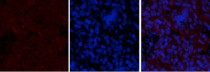
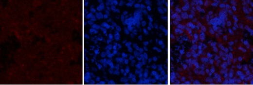
ARG66860 anti-GSK3 beta phospho (Ser9) antibody IHC-P image
Immunohistochemistry: Paraffin-embedded Rat spleen tissue stained with ARG66860 anti-GSK3 beta phospho (Ser9) antibody (red) at 1:200 dilution, overnight at 4°C. DAPI (blue) for nuclear staining.
-
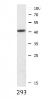
ARG66860 anti-GSK3 beta phospho (Ser9) antibody WB image
Western blot: 293 cell lysate stained with ARG66860 anti-GSK3 beta phospho (Ser9) antibody at 1:1000 dilution.
-

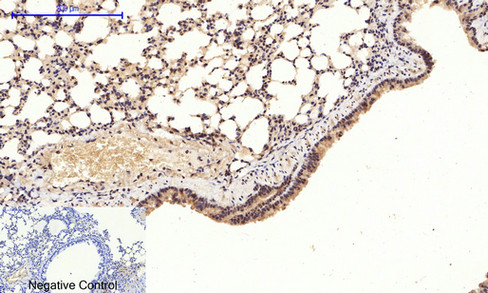
ARG66860 anti-GSK3 beta phospho (Ser9) antibody IHC-P image
Immunohistochemistry: Paraffin-embedded Mouse lung tissue. Antigen Retrieval: Boil tissue section in Sodium citrate buffer (pH 6.0) for 20 min. The tissue section was stained with ARG66860 anti-GSK3 beta phospho (Ser9) antibody at 1:200 dilution, overnight at 4°C. Negative control was used by secondary antibody only.
-
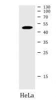
ARG66860 anti-GSK3 beta phospho (Ser9) antibody WB image
Western blot: HeLa cell lysate stained with ARG66860 anti-GSK3 beta phospho (Ser9) antibody at 1:1000 dilution.