ARG43428
anti-GSDMD antibody [6D11]
anti-GSDMD antibody [6D11] for Flow cytometry,ICC/IF,IHC-Formalin-fixed paraffin-embedded sections,Western blot and Human
Overview
| Product Description | Mouse Monoclonal antibody [6D11] recognizes GSDMD |
|---|---|
| Tested Reactivity | Hu |
| Tested Application | FACS, ICC/IF, IHC-P, WB |
| Host | Mouse |
| Clonality | Monoclonal |
| Clone | 6D11 |
| Isotype | IgG2b |
| Target Name | GSDMD |
| Antigen Species | Human |
| Immunogen | Recombinant protein corresponding to M1-H484 of Human GSDMD. |
| Conjugation | Un-conjugated |
| Alternate Names | FKSG10; DF5L; Gasdermin domain-containing protein 1; Gasdermin-D; DFNA5L; GSDMDC1 |
Application Instructions
| Application Suggestion |
|
||||||||||
|---|---|---|---|---|---|---|---|---|---|---|---|
| Application Note | IHC-P: Antigen Retrieval: Heat mediation was performed in EDTA buffer (pH 8.0). * The dilutions indicate recommended starting dilutions and the optimal dilutions or concentrations should be determined by the scientist. |
||||||||||
| Observed Size | ~ 53 kDa |
Properties
| Form | Liquid |
|---|---|
| Purification | Affinity purification with immunogen. |
| Buffer | 0.2% Na2HPO4, 0.9% NaCl and 4% Trehalose. |
| Stabilizer | 4% Trehalose |
| Concentration | 0.5 mg/ml |
| Storage Instruction | For continuous use, store undiluted antibody at 2-8°C for up to a week. For long-term storage, aliquot and store at -20°C or below. Storage in frost free freezers is not recommended. Avoid repeated freeze/thaw cycles. Suggest spin the vial prior to opening. The antibody solution should be gently mixed before use. |
| Note | For laboratory research only, not for drug, diagnostic or other use. |
Bioinformation
| Database Links | |
|---|---|
| Gene Symbol | GSDMD |
| Gene Full Name | gasdermin D |
| Background | Gasdermin D is a member of the gasdermin family. Members of this family appear to play a role in regulation of epithelial proliferation. Gasdermin D has been suggested to act as a tumor suppressor. Alternatively spliced transcript variants have been described. [provided by RefSeq, Oct 2009] |
| Function | [Gasdermin-D, N-terminal]: Promotes pyroptosis in response to microbial infection and danger signals. Produced by the cleavage of gasdermin-D by inflammatory caspases CASP1 or CASP4 in response to canonical, as well as non-canonical (such as cytosolic LPS) inflammasome activators (PubMed:26375003, PubMed:26375259, PubMed:27418190). After cleavage, moves to the plasma membrane where it strongly binds to inner leaflet lipids, including monophosphorylated phosphatidylinositols, such as phosphatidylinositol 4-phosphate, bisphosphorylated phosphatidylinositols, such as phosphatidylinositol (4,5)-bisphosphate, as well as phosphatidylinositol (3,4,5)-bisphosphate, and more weakly to phosphatidic acid and phosphatidylserine (PubMed:27281216). Homooligomerizes within the membrane and forms pores of 10 - 15 nanometers (nm) of inner diameter, possibly allowing the release of mature IL1B and triggering pyroptosis (PubMed:27418190, PubMed:27281216). Exhibits bactericidal activity. Gasdermin-D, N-terminal released from pyroptotic cells into the extracellular milieu rapidly binds to and kills both Gram-negative and Gram-positive bacteria, without harming neighboring mammalian cells, as it does not disrupt the plasma membrane from the outside due to lipid-binding specificity (PubMed:27281216). Under cell culture conditions, also active against intracellular bacteria, such as Listeria monocytogenes (By similarity). Strongly binds to bacterial and mitochondrial lipids, including cardiolipin. Does not bind to unphosphorylated phosphatidylinositol, phosphatidylethanolamine nor phosphatidylcholine (PubMed:27281216). [UniProt] |
| Cellular Localization | Gasdermin-D: Cytoplasm, cytosol. Inflammasome. Note=In response to a canonical inflammasome stimulus, such as nigericin, recruited to NLRP3 inflammasone with similar kinetics to that of uncleaved CASP1 precursor. Gasdermin-D, N-terminal: Cell membrane. Secreted. Note=Released in the extracellular milieu following pyroptosis. [UniProt] |
| Calculated MW | 53 kDa |
| PTM | Cleavage at Asp-275 by CASP1 (mature and uncleaved precursor forms) or CASP4 relieves autoinhibition and is sufficient to initiate pyroptosis. [UniProt] |
Images (6) Click the Picture to Zoom In
-
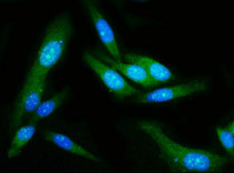
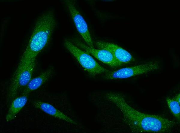
ARG43428 anti-GSDMD antibody [6D11] ICC/IF image
Immunofluorescence: PC-3 cells were blocked with 10% goat serum and then stained with ARG43428 anti-GSDMD antibody [6D11] (green) at 5 µg/ml dilution, overnight at 4°C. DAPI (blue) for nuclear staining.
-
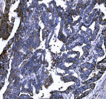
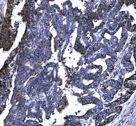
ARG43428 anti-GSDMD antibody [6D11] IHC-P image
Immunohistochemistry: Paraffin-embedded Human cervical cancer tissue. Antigen Retrieval: Heat mediation was performed in EDTA buffer (pH 8.0). The tissue section was blocked with 10% goat serum. The tissue section was then stained with ARG43428 anti-GSDMD antibody [6D11] at 2 µg/ml dilution, overnight at 4°C.
-
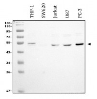
ARG43428 anti-GSDMD antibody [6D11] WB image
Western blot: 30 µg of sample under reducing conditions. THP-1, SW620, Jurkat, U87 and PC-3 whole cell lysates stained with ARG43428 anti-GSDMD antibody [6D11] at 0.5 µg/ml dilution, overnight at 4°C.
-
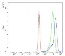
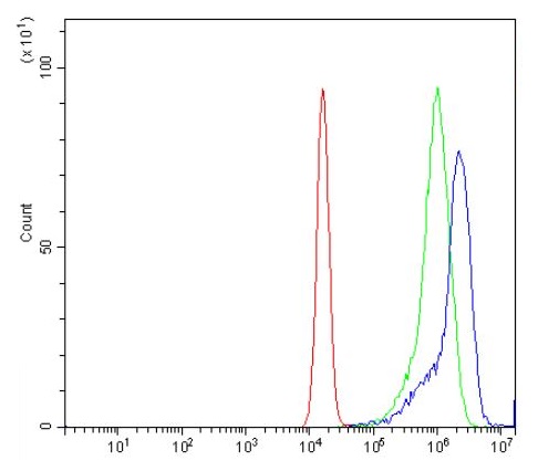
ARG43428 anti-GSDMD antibody [6D11] FACS image
Flow Cytometry: Jurkat cells were blocked with 10% normal goat serum and then stained with ARG43428 anti-GSDMD antibody [6D11] (blue) at 1 µg/10^6 cells for 30 min at 20°C, followed by incubation with DyLight®488 labelled secondary antibody. Isotype control antibody (green) was mouse IgG (1 µg/10^6 cells) used under the same conditions. Unlabelled sample (Red) was also used as a control.
-

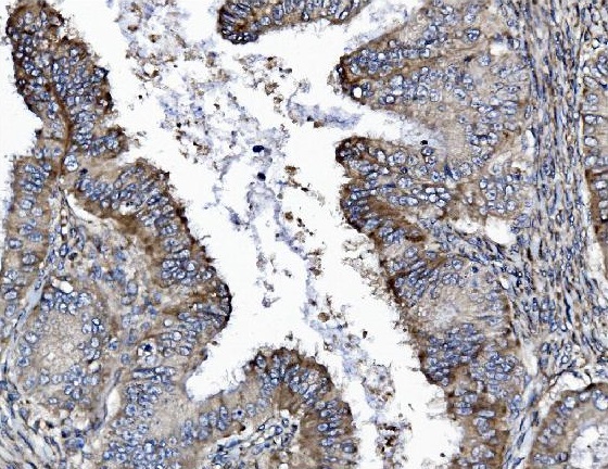
ARG43428 anti-GSDMD antibody [6D11] IHC-P image
Immunohistochemistry: Paraffin-embedded Human cervical intraepithelial neoplasia tissue. Antigen Retrieval: Heat mediation was performed in EDTA buffer (pH 8.0). The tissue section was blocked with 10% goat serum. The tissue section was then stained with ARG43428 anti-GSDMD antibody [6D11] at 2 µg/ml dilution, overnight at 4°C.
-
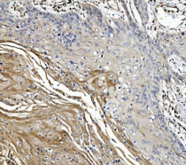
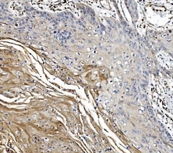
ARG43428 anti-GSDMD antibody [6D11] IHC-P image
Immunohistochemistry: Paraffin-embedded Human esophageal squamous carcinoma tissue. Antigen Retrieval: Heat mediation was performed in EDTA buffer (pH 8.0). The tissue section was blocked with 10% goat serum. The tissue section was then stained with ARG43428 anti-GSDMD antibody [6D11] at 2 µg/ml dilution, overnight at 4°C.