ARG63994
anti-GRP94 antibody
anti-GRP94 antibody for Flow cytometry,ICC/IF,IHC-Formalin-fixed paraffin-embedded sections,Western blot and Human,Mouse
Cancer antibody; Controls and Markers antibody; Metabolism antibody; Signaling Transduction antibody
Overview
| Product Description | Goat Polyclonal antibody recognizes GRP94 |
|---|---|
| Tested Reactivity | Hu, Ms |
| Predict Reactivity | Cow, Rat, Dog, Pig, Zfsh |
| Tested Application | FACS, ICC/IF, IHC-P, WB |
| Host | Goat |
| Clonality | Polyclonal |
| Isotype | IgG |
| Target Name | GRP94 |
| Antigen Species | Human |
| Immunogen | C-KEGVKFDESEKTKE |
| Conjugation | Un-conjugated |
| Alternate Names | Tumor rejection antigen 1; gp96 homolog; Endoplasmin; 94 kDa glucose-regulated protein; HEL-S-125m; GP96; GRP94; TRA1; HEL35; GRP-94; Heat shock protein 90 kDa beta member 1; ECGP |
Application Instructions
| Application Suggestion |
|
||||||||||
|---|---|---|---|---|---|---|---|---|---|---|---|
| Application Note | WB: Recommend incubate at RT for 1h. IHC-P: Antigen Retrieval: Steam tissue section in Citrate buffer (pH 6.0). * The dilutions indicate recommended starting dilutions and the optimal dilutions or concentrations should be determined by the scientist. |
Properties
| Form | Liquid |
|---|---|
| Purification | Purified from goat serum by ammonium sulphate precipitation followed by antigen affinity chromatography using the immunizing peptide. |
| Buffer | Tris saline (pH 7.3), 0.02% Sodium azide and 0.5% BSA |
| Preservative | 0.02% Sodium azide |
| Stabilizer | 0.5% BSA |
| Concentration | 0.5 mg/ml |
| Storage Instruction | For continuous use, store undiluted antibody at 2-8°C for up to a week. For long-term storage, aliquot and store at -20°C or below. Storage in frost free freezers is not recommended. Avoid repeated freeze/thaw cycles. Suggest spin the vial prior to opening. The antibody solution should be gently mixed before use. |
| Note | For laboratory research only, not for drug, diagnostic or other use. |
Bioinformation
| Database Links | |
|---|---|
| Background | This gene encodes a member of a family of adenosine triphosphate(ATP)-metabolizing molecular chaperones with roles in stabilizing and folding other proteins. The encoded protein is localized to melanosomes and the endoplasmic reticulum. Expression of this protein is associated with a variety of pathogenic states, including tumor formation. There is a microRNA gene located within the 5' exon of this gene. [provided by RefSeq, Aug 2012] |
| Research Area | Cancer antibody; Controls and Markers antibody; Metabolism antibody; Signaling Transduction antibody |
| Calculated MW | 92 kDa |
Images (8) Click the Picture to Zoom In
-
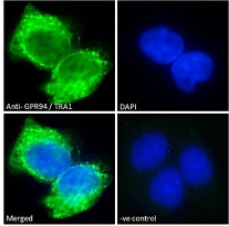
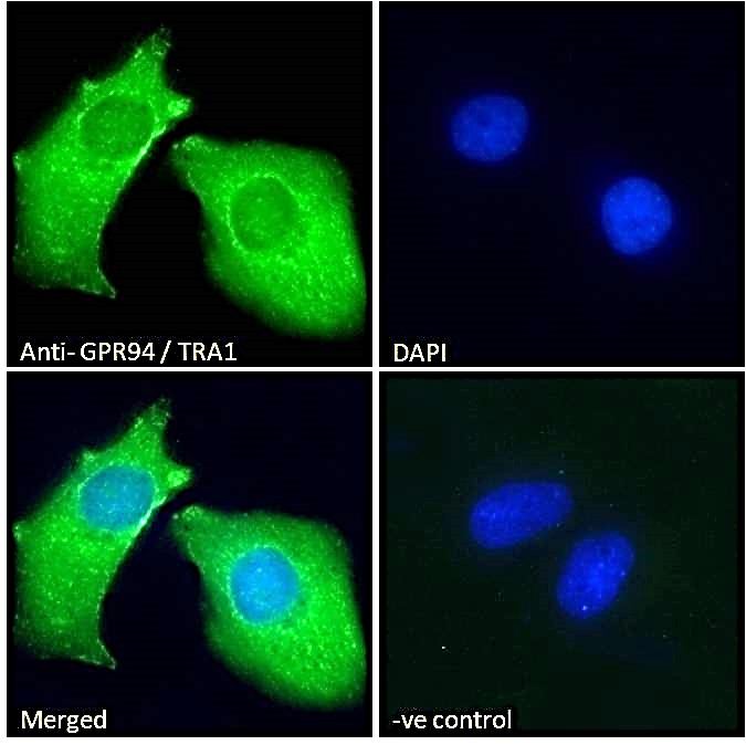
ARG63994 anti-GRP94 antibody ICC/IF image
Immunofluorescence: Paraformaldehyde fixed A431 cells permeabilized with 0.15% Triton. Cells were stained with ARG63994 anti-GRP94 antibody (green) at 10 µg/ml dilution for 1 hour. DAPI (blue) for nuclear staining. Negative control: Unimmunized goat IgG (green) at 10 µg/ml dilution.
-
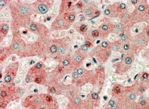
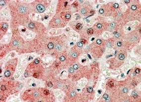
ARG63994 anti-GRP94 antibody IHC-P image
Immunohistochemistry: Paraffin-embedded Human liver tissue. Antigen Retrieval: Steam tissue section in Citrate buffer (pH 6.0). The tissue section was stained with ARG63994 anti-GRP94 antibody at 3.8 µg/ml dilution followed by AP-staining.
-
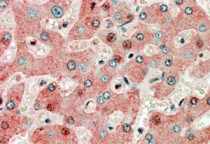
ARG63994 anti-GRP94 antibody IHC-P image
Immunohistochemistry: Paraffin embedded Human Liver. (Steamed antigen retrieval with citrate buffer pH 6) stained with ARG63994 anti-GRP94 antibody at 3.8 µg/ml dilution followed by AP-staining.
-
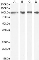
ARG63994 anti-GRP94 antibody WB image
Western blot: 35 µg of U2OS (A), HEK293 (B), HeLa (C) and A549 (D) cell lysates (in RIPA buffer) stained with ARG63994 anti-GRP94 antibody at 0.1 µg/ml (A-C) and 0.3 µg/ml (D) dilutions and incubated at RT for 1 hour.
-
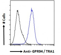
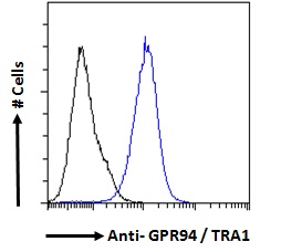
ARG63994 anti-GRP94 antibody FACS image
Flow Cytometry: Paraformaldehyde-fixed A431 cells permeabilized with 0.5% Triton. Cells were stained with ARG63994 anti-GRP94 antibody (blue line) at 10 µg/ml dilution for 1 hour, followed by incubation with Alexa FluorR 488 labelled secondary antibody. IgG control: Unimmunized goat IgG (black line).
-
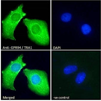
ARG63994 anti-GRP94 antibody ICC/IF image
Immunofluorescence: Paraformaldehyde fixed HeLa cells permeabilized with 0.15% Triton. Cells were stained with ARG63994 anti-GRP94 antibody (green) at 10 µg/ml dilution for 1 hour. DAPI (blue) for nuclear staining. Negative control: Unimmunized goat IgG (green) at 10 µg/ml dilution.
-

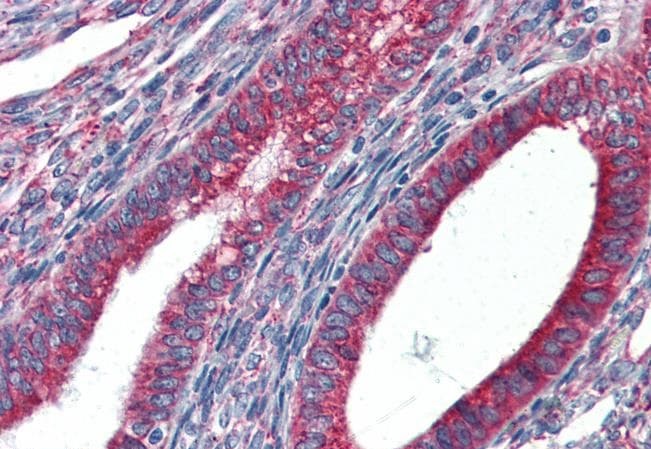
ARG63994 anti-GRP94 antibody IHC-P image
Immunohistochemistry: Paraffin-embedded Human uterus tissue. Antigen Retrieval: Steam tissue section in Citrate buffer (pH 6.0). The tissue section was stained with ARG63994 anti-GRP94 antibody at 5 µg/ml dilution followed by AP-staining.
-
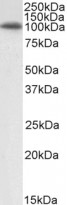
ARG63994 anti-GRP94 antibody WB image
Western blot: 35 µg of NIH/3T3 cell lysate (in RIPA buffer) stained with ARG63994 anti-GRP94 antibody at 0.1 µg/ml dilution and incubated at RT for 1 hour.