ARG59676
anti-GRIK1 / GluR5 antibody
anti-GRIK1 / GluR5 antibody for IHC-Formalin-fixed paraffin-embedded sections,Western blot and Human,Mouse,Rat
Overview
| Product Description | Rabbit Polyclonal antibody recognizes GRIK1 / GluR5 |
|---|---|
| Tested Reactivity | Hu, Ms, Rat |
| Tested Application | IHC-P, WB |
| Host | Rabbit |
| Clonality | Polyclonal |
| Isotype | IgG |
| Target Name | GRIK1 / GluR5 |
| Antigen Species | Human |
| Immunogen | Recombinant protein corresponding to R271-I450 of Human GRIK1. |
| Conjugation | Un-conjugated |
| Alternate Names | GluR5; GluK1; GLUR5; EEA3; GluR-5; Excitatory amino acid receptor 3; Glutamate receptor ionotropic, kainate 1; EAA3; Glutamate receptor 5; GLR5 |
Application Instructions
| Application Suggestion |
|
||||||
|---|---|---|---|---|---|---|---|
| Application Note | IHC-P: Antigen Retrieval: Heat mediation was performed in Citrate buffer (pH 6.0) for 20 min. * The dilutions indicate recommended starting dilutions and the optimal dilutions or concentrations should be determined by the scientist. |
Properties
| Form | Liquid |
|---|---|
| Purification | Affinity purification with immunogen. |
| Buffer | 0.9% NaCl, 0.2% Na2HPO4, 0.05% Sodium azide and 5% BSA. |
| Preservative | 0.05% Sodium azide |
| Stabilizer | 5% BSA |
| Concentration | 0.5 mg/ml |
| Storage Instruction | For continuous use, store undiluted antibody at 2-8°C for up to a week. For long-term storage, aliquot and store at -20°C or below. Storage in frost free freezers is not recommended. Avoid repeated freeze/thaw cycles. Suggest spin the vial prior to opening. The antibody solution should be gently mixed before use. |
| Note | For laboratory research only, not for drug, diagnostic or other use. |
Bioinformation
| Database Links |
Swiss-port # P22756 Rat Glutamate receptor ionotropic, kainate 1 Swiss-port # P39086 Human Glutamate receptor ionotropic, kainate 1 |
|---|---|
| Gene Symbol | GRIK1 |
| Gene Full Name | glutamate receptor, ionotropic, kainate 1 |
| Background | Glutamate receptors are the predominant excitatory neurotransmitter receptors in the mammalian brain and are activated in a variety of normal neurophysiologic processes. This gene product belongs to the kainate family of glutamate receptors, which are composed of four subunits and function as ligand-activated ion channels. The subunit encoded by this gene is subject to RNA editing (CAG->CGG; Q->R) within the second transmembrane domain, which is thought to alter the properties of ion flow. Alternative splicing, resulting in transcript variants encoding different isoforms, has been noted for this gene. [provided by RefSeq, Jul 2008] |
| Function | Ionotropic glutamate receptor. L-glutamate acts as an excitatory neurotransmitter at many synapses in the central nervous system. Binding of the excitatory neurotransmitter L-glutamate induces a conformation change, leading to the opening of the cation channel, and thereby converts the chemical signal to an electrical impulse. The receptor then desensitizes rapidly and enters a transient inactive state, characterized by the presence of bound agonist. May be involved in the transmission of light information from the retina to the hypothalamus. [UniProt] |
| Cellular Localization | Cell membrane; Multi-pass membrane protein. Cell junction, synapse, postsynaptic cell membrane; Multi-pass membrane protein. [UniProt] |
| Calculated MW | 104 kDa |
Images (7) Click the Picture to Zoom In
-
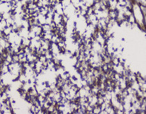
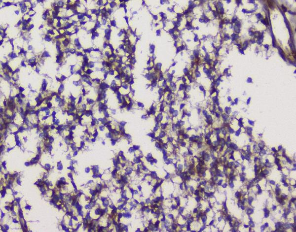
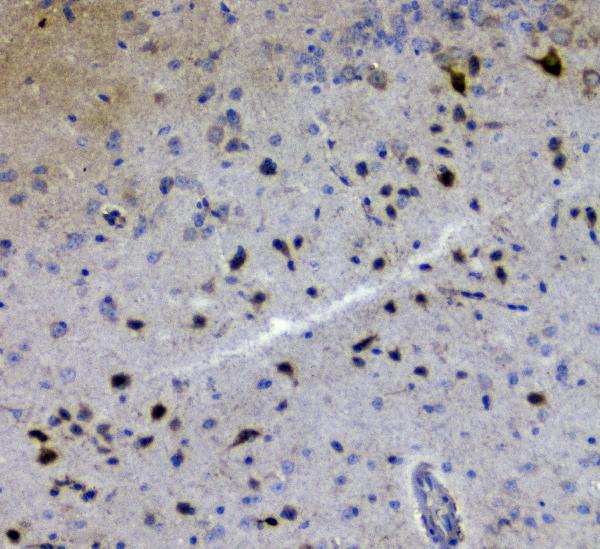
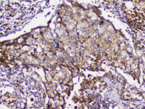
ARG59676 anti-GRIK1 / GluR5 antibody IHC-P image
Immunohistochemistry: Paraffin-embedded Human glioma tissue. Antigen Retrieval: Heat mediation was performed in Citrate buffer (pH 6.0) for 20 min. The tissue section was blocked with 10% goat serum. The tissue section was then stained with ARG59676 anti-GRIK1 / GluR5 antibody at 1 µg/ml dilution, overnight at 4°C.
-

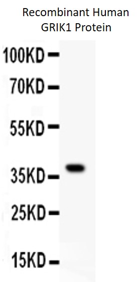
ARG59676 anti-GRIK1 / GluR5 antibody WB image
Western blot: 0.5 ng of Recombinant Human GRIK1 Protein stained with ARG59676 anti-GRIK1 / GluR5 antibody at 0.5 µg/ml dilution.
-
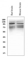
ARG59676 anti-GRIK1 / GluR5 antibody WB image
Western blot: 50 µg of sample under reducing conditions. Rat brain and Mouse brain lysates stained with ARG59676 anti-GRIK1 / GluR5 antibody at 0.5 µg/ml dilution, overnight at 4°C.
-
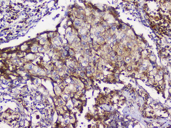
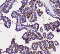
ARG59676 anti-GRIK1 / GluR5 antibody IHC-P image
Immunohistochemistry: Paraffin-embedded Human lung cancer tissue. Antigen Retrieval: Heat mediation was performed in Citrate buffer (pH 6.0) for 20 min. The tissue section was blocked with 10% goat serum. The tissue section was then stained with ARG59676 anti-GRIK1 / GluR5 antibody at 1 µg/ml dilution, overnight at 4°C.
-
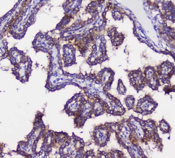
ARG59676 anti-GRIK1 / GluR5 antibody IHC-P image
Immunohistochemistry: Paraffin-embedded Human thyroid cancer tissue. Antigen Retrieval: Heat mediation was performed in Citrate buffer (pH 6.0) for 20 min. The tissue section was blocked with 10% goat serum. The tissue section was then stained with ARG59676 anti-GRIK1 / GluR5 antibody at 1 µg/ml dilution, overnight at 4°C.
-

ARG59676 anti-GRIK1 / GluR5 antibody IHC-P image
Immunohistochemistry: Paraffin-embedded Mouse brain tissue tissue. Antigen Retrieval: Heat mediation was performed in Citrate buffer (pH 6.0) for 20 min. The tissue section was blocked with 10% goat serum. The tissue section was then stained with ARG59676 anti-GRIK1 / GluR5 antibody at 1 µg/ml dilution, overnight at 4°C.
-
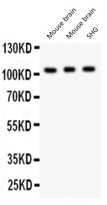
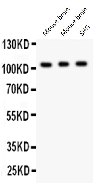
ARG59676 anti-GRIK1 / GluR5 antibody WB image
Western blot: 50 µg of Mouse brain (Lane 1, 2) and 40 µg of SHG whole cell lysate (Lane 3) stained with ARG59676 anti-GRIK1 / GluR5 antibody at 0.5 µg/ml dilution.