ARG42840
anti-GRB10 antibody
anti-GRB10 antibody for Flow cytometry,IHC-Formalin-fixed paraffin-embedded sections,Western blot and Human,Mouse,Rat
Overview
| Product Description | Rabbit Polyclonal antibody recognizes GRB10 |
|---|---|
| Tested Reactivity | Hu, Ms, Rat |
| Tested Application | FACS, IHC-P, WB |
| Host | Rabbit |
| Clonality | Polyclonal |
| Isotype | IgG |
| Target Name | GRB10 |
| Antigen Species | Human |
| Immunogen | Recombinant protein corresponding to M1-K251 of Human GRB10. |
| Conjugation | Un-conjugated |
| Alternate Names | GRB10 adapter protein; GRB-IR; Insulin receptor-binding protein Grb-IR; Grb-10; MEG1; RSS; Growth factor receptor-bound protein 10; IRBP |
Application Instructions
| Application Suggestion |
|
||||||||
|---|---|---|---|---|---|---|---|---|---|
| Application Note | IHC-P: Antigen Retrieval: Heat mediation was performed in EDTA buffer (pH 8.0). * The dilutions indicate recommended starting dilutions and the optimal dilutions or concentrations should be determined by the scientist. |
||||||||
| Observed Size | ~ 75 kDa |
Properties
| Form | Liquid |
|---|---|
| Purification | Affinity purification with immunogen. |
| Buffer | 0.2% Na2HPO4, 0.9% NaCl, 0.01% Sodium azide and 4% Trehalose. |
| Preservative | 0.01% Sodium azide |
| Stabilizer | 4% Trehalose |
| Concentration | 0.5 mg/ml |
| Storage Instruction | For continuous use, store undiluted antibody at 2-8°C for up to a week. For long-term storage, aliquot and store at -20°C or below. Storage in frost free freezers is not recommended. Avoid repeated freeze/thaw cycles. Suggest spin the vial prior to opening. The antibody solution should be gently mixed before use. |
| Note | For laboratory research only, not for drug, diagnostic or other use. |
Bioinformation
| Database Links | |
|---|---|
| Gene Symbol | GRB10 |
| Gene Full Name | growth factor receptor-bound protein 10 |
| Background | The product of this gene belongs to a small family of adapter proteins that are known to interact with a number of receptor tyrosine kinases and signaling molecules. This gene encodes a growth factor receptor-binding protein that interacts with insulin receptors and insulin-like growth-factor receptors. Overexpression of some isoforms of the encoded protein inhibits tyrosine kinase activity and results in growth suppression. This gene is imprinted in a highly isoform- and tissue-specific manner, with expression observed from the paternal allele in the brain, and from the maternal allele in the placental trophoblasts. Alternatively spliced transcript variants encoding different isoforms have been identified. [provided by RefSeq, Oct 2010] |
| Function | Adapter protein which modulates coupling of a number of cell surface receptor kinases with specific signaling pathways. Binds to, and suppress signals from, activated receptors tyrosine kinases, including the insulin (INSR) and insulin-like growth factor (IGF1R) receptors. The inhibitory effect can be achieved by 2 mechanisms: interference with the signaling pathway and increased receptor degradation. Delays and reduces AKT1 phosphorylation in response to insulin stimulation. Blocks association between INSR and IRS1 and IRS2 and prevents insulin-stimulated IRS1 and IRS2 tyrosine phosphorylation. Recruits NEDD4 to IGF1R, leading to IGF1R ubiquitination, increased internalization and degradation by both the proteasomal and lysosomal pathways. May play a role in mediating insulin-stimulated ubiquitination of INSR, leading to proteasomal degradation. Negatively regulates Wnt signaling by interacting with LRP6 intracellular portion and interfering with the binding of AXIN1 to LRP6. Positive regulator of the KDR/VEGFR-2 signaling pathway. May inhibit NEDD4-mediated degradation of KDR/VEGFR-2. [UniProt] |
| Cellular Localization | Cytoplasm. Note=When complexed with NEDD4 and IGF1R, follows IGF1R internalization, remaining associated with early endosomes. Uncouples from IGF1R-containing endosomes before the sorting of the receptor to the lysosomal compartment (By similarity). [UniProt] |
| Calculated MW | 67 kDa |
| PTM | Phosphorylated on serine residues upon EGF, FGF and PDGF stimulation (By similarity). Phosphorylated at Tyr-67 by TEC. [UniProt] |
Images (6) Click the Picture to Zoom In
-
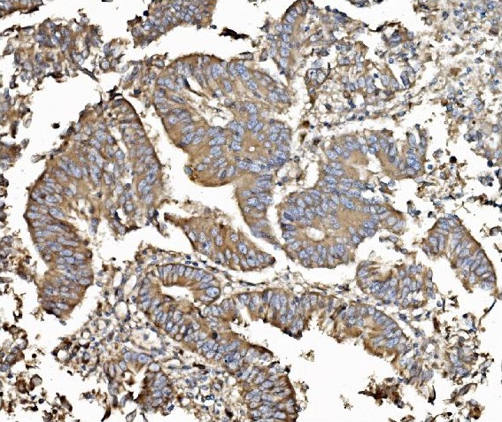
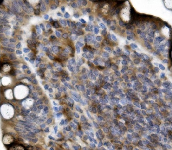
ARG42840 anti-GRB10 antibody IHC-P image
Immunohistochemistry: Paraffin-embedded Human rectal cancer tissue. Antigen Retrieval: Heat mediation was performed in EDTA buffer (pH 8.0). The tissue section was blocked with 10% goat serum. The tissue section was then stained with ARG42840 anti-GRB10 antibody at 2 µg/ml dilution, overnight at 4°C.
-
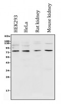
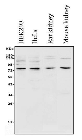
ARG42840 anti-GRB10 antibody WB image
Western blot: 50 µg of sample under reducing conditions. HEK293, HeLa, Rat kidney and Mouse kidney lysates stained with ARG42840 anti-GRB10 antibody at 0.5 µg/ml dilution, overnight at 4°C.
-
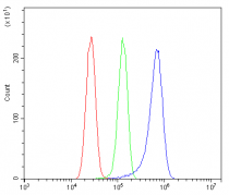
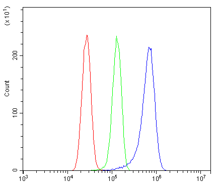
ARG42840 anti-GRB10 antibody FACS image
Flow Cytometry: U-87 MG cells were blocked with 10% normal goat serum and then stained with ARG42840 anti-GRB10 antibody (blue) at 1 µg/10^6 cells for 30 min at 20°C, followed by incubation with DyLight®488 labelled secondary antibody. Isotype control antibody (green) was Rabbit IgG (1 µg/10^6 cells) used under the same conditions. Unlabelled sample (red) was also used as a control.
-
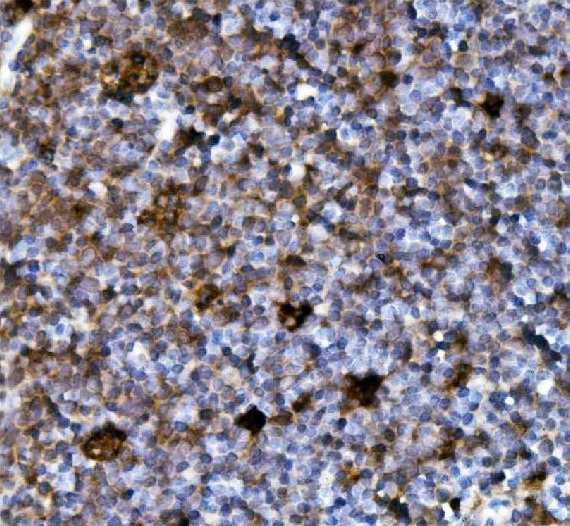
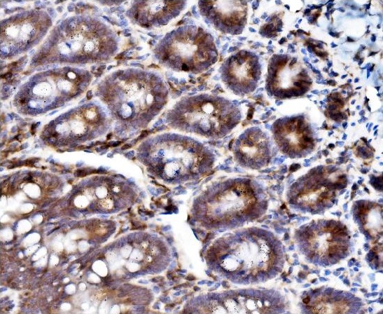
ARG42840 anti-GRB10 antibody IHC-P image
Immunohistochemistry: Paraffin-embedded Mouse intestine tissue. Antigen Retrieval: Heat mediation was performed in EDTA buffer (pH 8.0). The tissue section was blocked with 10% goat serum. The tissue section was then stained with ARG42840 anti-GRB10 antibody at 2 µg/ml dilution, overnight at 4°C.
-

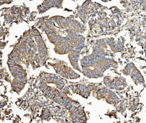
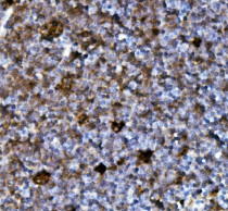
ARG42840 anti-GRB10 antibody IHC-P image
Immunohistochemistry: Paraffin-embedded Rat intestine tissue. Antigen Retrieval: Heat mediation was performed in EDTA buffer (pH 8.0). The tissue section was blocked with 10% goat serum. The tissue section was then stained with ARG42840 anti-GRB10 antibody at 2 µg/ml dilution, overnight at 4°C.
-

ARG42840 anti-GRB10 antibody IHC-P image
Immunohistochemistry: Paraffin-embedded Rat intestine tissue. Antigen Retrieval: Heat mediation was performed in EDTA buffer (pH 8.0). The tissue section was blocked with 10% goat serum. The tissue section was then stained with ARG42840 anti-GRB10 antibody at 2 µg/ml dilution, overnight at 4°C.