ARG45514
anti-GPR56 antibody
anti-GPR56 antibody for Flow cytometry,IHC-Formalin-fixed paraffin-embedded sections,Western blot and Mouse,Rat
Overview
| Product Description | Rabbit Polyclonal antibody recognizes GPR56 |
|---|---|
| Tested Reactivity | Ms, Rat |
| Tested Application | FACS, IHC-P, WB |
| Host | Rabbit |
| Clonality | Polyclonal |
| Isotype | IgG |
| Target Name | GPR56 |
| Antigen Species | Mouse |
| Immunogen | Recombinant protein containing to mouse GPR56. |
| Conjugation | Un-conjugated |
| Alternate Names | ADGRG1; adhesion G protein-coupled receptor G1; BPPR; G-protein coupled receptor 56; C; GPR56 subunit alpha; BFPP; GPR56 NT; Protein TM7XN1; TM7XN1; GPR56 seven-transmembrane subunit; GPR56 extracellular subunit; N; GPR56 subunit beta; GPR56 7TM; GPR56; TM7LN4; GPR56 CT |
Application Instructions
| Application Suggestion |
|
||||||||
|---|---|---|---|---|---|---|---|---|---|
| Application Note | * The dilutions indicate recommended starting dilutions and the optimal dilutions or concentrations should be determined by the scientist. |
Properties
| Form | Liquid |
|---|---|
| Purification | Affinity purified |
| Buffer | 0.2% Na2HPO4, 0.9% NaCl and 4% Trehalose. |
| Stabilizer | 4% Trehalose |
| Concentration | 0.5 mg/ml |
| Storage Instruction | For continuous use, store undiluted antibody at 2-8°C for up to a week. For long-term storage, aliquot and store at -20°C or below. Storage in frost free freezers is not recommended. Avoid repeated freeze/thaw cycles. Suggest spin the vial prior to opening. The antibody solution should be gently mixed before use. |
| Note | For laboratory research only, not for drug, diagnostic or other use. |
Bioinformation
| Gene Symbol | ADGRG1 |
|---|---|
| Gene Full Name | adhesion G protein-coupled receptor G1 |
| Background | This gene encodes a member of the G protein-coupled receptor family and regulates brain cortical patterning. The encoded protein binds specifically to transglutaminase 2, a component of tissue and tumor stroma implicated as an inhibitor of tumor progression. Mutations in this gene are associated with a brain malformation known as bilateral frontoparietal polymicrogyria. Alternative splicing results in multiple transcript variants. [provided by RefSeq, Feb 2014] |
| Function | Receptor involved in cell adhesion and probably in cell-cell interactions. Mediates cell matrix adhesion in developing neurons and hematopoietic stem cells. Receptor for collagen III/COL3A1 in the developing brain and involved in regulation of cortical development, specifically in maintenance of the pial basement membrane integrity and in cortical lamination (By similarity). Binding to the COL3A1 ligand inhibits neuronal migration and activates the RhoA pathway by coupling to GNA13 and possibly GNA12 (PubMed:22238662). Plays a role in the maintenance of hematopoietic stem cells and/or leukemia stem cells in bone marrow niche (By similarity). Plays a critical role in cancer progression by inhibiting VEGFA production threreby inhibiting angiogenesis through a signaling pathway mediated by PRKCA (PubMed:16757564, PubMed:21724588). Plays an essential role in testis development (By similarity). [ADGRG1 N-terminal fragment]: Plays a critical role in cancer progression by activating VEGFA production and angiogenesis through a signaling pathway mediated by PRKCA (PubMed:21724588). [UniProt] |
| Cellular Localization | Cell membrane; Multi-pass membrane protein. ADGRG1 N-terminal fragment: Secreted. ADGRG1 C-terminal fragment: Membrane raft. Note=Interaction with its ligand COL3A1 leads to the release of ADGRG1 NT from the membrane and triggers the association of ADGRG1 CT with lipid rafts. [UniProt] |
| Calculated MW | 78 kDa |
| PTM | Autoproteolytically cleaved into 2 fragments; the large extracellular N-terminal fragment (ADGRG1 NT) and the membrane-bound C-terminal fragment (ADGRG1 CT) predominantly remain associated and non-covalently linked. Shedding to yield the secreted ADGRG1 N-terminal fragment seems to involve metalloprotease(s) (PubMed:22333914). N-glycosylated. Contains sialic acid residues. Ubiquitinated. Undergoes polyubiquitination upon activation. [UniProt] |
Images (5) Click the Picture to Zoom In
-

ARG45514 anti-GPR56 antibody WB image
Western blot: Mouse heart stained with ARG45514 anti-GPR56 antibody at 0.5 μg/ml dilution.
-

ARG45514 anti-GPR56 antibody FACS image
Flow Cytometry: RAW264.7 stained with ARG45514 anti-GPR56 antibody at 1 µg/10^6 cells dilution.
-
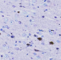
ARG45514 anti-GPR56 antibody IHC-P image
Immunohistochemistry: Rat brain stained with ARG45514 anti-GPR56 antibody at 2 μg/ml dilution.
-

ARG45514 anti-GPR56 antibody WB image
Western blot: Rat heart stained with ARG45514 anti-GPR56 antibody at 0.5 μg/ml dilution.
-
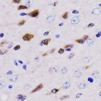
ARG45514 anti-GPR56 antibody IHC-P image
Immunohistochemistry: Mouse brain stained with ARG45514 anti-GPR56 antibody at 2 μg/ml dilution.


