ARG59645
anti-GPM6A antibody
anti-GPM6A antibody for ICC/IF,Western blot and Human,Rat
Overview
| Product Description | Rabbit Polyclonal antibody recognizes GPM6A |
|---|---|
| Tested Reactivity | Hu, Rat |
| Predict Reactivity | Ms, Cow, Dog, Gpig, Hrs, Rb, Zfsh |
| Tested Application | ICC/IF, WB |
| Host | Rabbit |
| Clonality | Polyclonal |
| Isotype | IgG |
| Target Name | GPM6A |
| Antigen Species | Human |
| Immunogen | Synthetic peptide around the C-terminal region of Human GPM6A. (within the following region: IAMVHYLMVLSANWAYVKDACRMQKYEDIKSKEEQELHDIHSTRSKERLN) |
| Conjugation | Un-conjugated |
| Alternate Names | M6A; Neuronal membrane glycoprotein M6-a; GPM6; M6a |
Application Instructions
| Predict Reactivity Note | Predicted Homology Based On Immunogen Sequence: Cow: 100%; Dog: 100%; Guinea Pig: 100%; Horse: 100%; Mouse: 100%; Rabbit: 100%; Zebrafish: 100% | ||||||
|---|---|---|---|---|---|---|---|
| Application Suggestion |
|
||||||
| Application Note | * The dilutions indicate recommended starting dilutions and the optimal dilutions or concentrations should be determined by the scientist. |
Properties
| Form | Liquid |
|---|---|
| Purification | Affinity purified. |
| Buffer | PBS, 0.09% (w/v) Sodium azide and 2% Sucrose. |
| Preservative | 0.09% (w/v) Sodium azide |
| Stabilizer | 2% Sucrose |
| Concentration | Batch dependent: 0.5 - 1 mg/ml |
| Storage Instruction | For continuous use, store undiluted antibody at 2-8°C for up to a week. For long-term storage, aliquot and store at -20°C or below. Storage in frost free freezers is not recommended. Avoid repeated freeze/thaw cycles. Suggest spin the vial prior to opening. The antibody solution should be gently mixed before use. |
| Note | For laboratory research only, not for drug, diagnostic or other use. |
Bioinformation
| Database Links |
Swiss-port # P51674 Human Neuronal membrane glycoprotein M6-a |
|---|---|
| Gene Symbol | GPM6A |
| Gene Full Name | glycoprotein M6A |
| Function | Involved in neuronal differentiation, including differentiation and migration of neuronal stem cells. Plays a role in neuronal plasticity and is involved in neurite and filopodia outgrowth, filopodia motility and probably synapse formation. GPM6A-induced filopodia formation involves mitogen-activated protein kinase (MAPK) and Src signaling pathways. May be involved in neuronal NGF-dependent Ca(2+) influx. May be involved in regulation of endocytosis and intracellular trafficking of G-protein-coupled receptors (GPCRs); enhances internalization and recycling of mu-type opioid receptor. [UniProt] |
| Cellular Localization | Cell membrane; Multi-pass membrane protein. Cell projection, axon. Cell projection, dendritic spine. Cell projection, filopodium. Note=Localizes to cholesterol-rich lipid rafts of the plasma membrane of hippocampal neurons. Localized to plasma membrane of cell bodies and neurites of hippocampal neurons. Localized in membrane protrusions (filopodia and spines) of primary hippocampal neurons. Localized to the growth cone edge membrane of elongating axons (By similarity). [UniProt] |
| Calculated MW | 31 kDa |
Images (4) Click the Picture to Zoom In
-

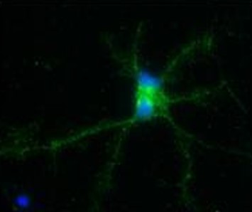
ARG59645 anti-GPM6A antibody ICC/IF image
Immunofluorescence: Rat hippocampal culture neurons stained with ARG59645 anti-GPM6A antibody (green) at 1:250 dilution. DAPI (blue) for nuclear staining.
-
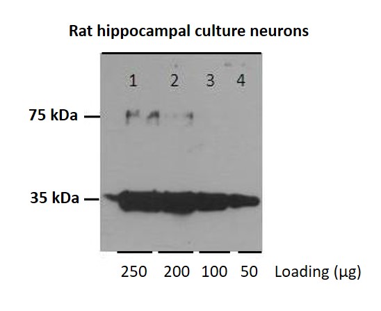
ARG59645 anti-GPM6A antibody WB image
Western blot: 250 µg, 200 µg, 100 µg and 50 µg of Rat hippocampal culture neurons stained with ARG59645 anti-GPM6A antibody at 1:1000 dilution.
-
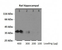
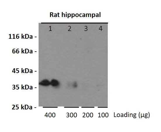
ARG59645 anti-GPM6A antibody WB image
Western blot: 400 µg, 300 µg, 200 µg and 100 µg of Rat hippocampal stained with ARG59645 anti-GPM6A antibody at 1:1000 dilution.
-
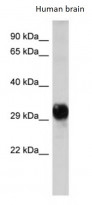
ARG59645 anti-GPM6A antibody WB image
Western blot: Human brain lysates stained with ARG59645 anti-GPM6A antibody at 1.0 µg/ml dilution.