ARG55094
anti-GOLPH3 antibody
anti-GOLPH3 antibody for Flow cytometry,IHC-Formalin-fixed paraffin-embedded sections,Western blot and Human
Controls and Markers antibody; Signaling Transduction antibody
Overview
| Product Description | Rabbit Polyclonal antibody recognizes GOLPH3 |
|---|---|
| Tested Reactivity | Hu |
| Predict Reactivity | Ms, Rat |
| Tested Application | FACS, IHC-P, WB |
| Host | Rabbit |
| Clonality | Polyclonal |
| Isotype | IgG |
| Target Name | GOLPH3 |
| Antigen Species | Human |
| Immunogen | KLH-conjugated synthetic peptide corresponding to aa. 1-30 (N-terminus) of Human GOLPH3. |
| Conjugation | Un-conjugated |
| Alternate Names | Mitochondrial DNA absence factor; Golgi phosphoprotein 3; GOPP1; Coat protein GPP34; MIDAS; Vps74; GPP34 |
Application Instructions
| Application Suggestion |
|
||||||||
|---|---|---|---|---|---|---|---|---|---|
| Application Note | * The dilutions indicate recommended starting dilutions and the optimal dilutions or concentrations should be determined by the scientist. | ||||||||
| Positive Control | HeLa |
Properties
| Form | Liquid |
|---|---|
| Purification | Saturated Ammonium Sulfate (SAS) precipitation followed by dialysis against PBS. |
| Buffer | PBS and 0.09% (W/V) Sodium azide |
| Preservative | 0.09% (W/V) Sodium azide |
| Storage Instruction | For continuous use, store undiluted antibody at 2-8°C for up to a week. For long-term storage, aliquot and store at -20°C or below. Storage in frost free freezers is not recommended. Avoid repeated freeze/thaw cycles. Suggest spin the vial prior to opening. The antibody solution should be gently mixed before use. |
| Note | For laboratory research only, not for drug, diagnostic or other use. |
Bioinformation
| Database Links | |
|---|---|
| Gene Symbol | GOLPH3 |
| Gene Full Name | golgi phosphoprotein 3 (coat-protein) |
| Background | The Golgi complex plays a key role in the sorting and modification of proteins exported from the endoplasmic reticulum. The protein encoded by this gene is a peripheral membrane protein of the Golgi stack and may have a regulatory role in Golgi trafficking. Several alternatively spliced transcript variants of this gene have been described, but the full-length nature of these variants has not been determined. [provided by RefSeq, Jul 2008] |
| Function | Phosphatidylinositol-4-phosphate-binding protein that links Golgi membranes to the cytoskeleton and may participate in the tensile force required for vesicle budding from the Golgi. Thereby, may play a role in Golgi membrane trafficking and could indirectly give its flattened shape to the Golgi apparatus. May also bind to the coatomer to regulate Golgi membrane trafficking. May play a role in anterograde transport from the Golgi to the plasma membrane and regulate secretion. Has also been involved in the control of the localization of Golgi enzymes through interaction with their cytoplasmic part. May play an indirect role in cell migration. Has also been involved in the modulation of mTOR signaling. May also be involved in the regulation of mitochondrial lipids biosynthesis. [UniProt] |
| Cellular Localization | Golgi apparatus, Golgi stack membrane; Peripheral membrane protein; Cytoplasmic side. Golgi apparatus, trans-Golgi network membrane; Peripheral membrane protein; Cytoplasmic side. Mitochondrion intermembrane space. Cell membrane. Endosome. Note=Phosphatidylinositol 4-phosphate-binding and oligomerization participate in the recruitment onto Golgi membranes. |
| Research Area | Controls and Markers antibody; Signaling Transduction antibody |
| Calculated MW | 34 kDa |
| PTM | Phosphorylated. |
Images (3) Click the Picture to Zoom In
-
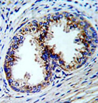
ARG55094 anti-GOLPH3 antibody IHC-P image
Immunohistochemistry: Formalin-fixed and paraffin-embedded Human prostate carcinoma stained with ARG55094 anti-GOLPH3 antibody.
-

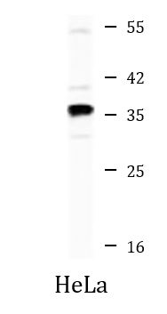
ARG55094 anti-GOLPH3 antibody WB image
Western blot: 35 µg of HeLa cell lysate stained with ARG55094 anti-GOLPH3 antibody.
-
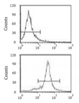
ARG55094 anti-GOLPH3 antibody FACS image
Flow Cytometry: HeLa cells stained with ARG55094 anti-GOLPH3 antibody (bottom histogram) or without primary antibody control (top histogram), followed by incubation with FITC labelled secondary antibody.