ARG43292
anti-GNAS antibody
anti-GNAS antibody for ICC/IF,Immunoprecipitation,Western blot and Human,Mouse,Rat
Overview
| Product Description | Rabbit Polyclonal antibody recognizes GNAS |
|---|---|
| Tested Reactivity | Hu, Ms, Rat |
| Tested Application | ICC/IF, IP, WB |
| Host | Rabbit |
| Clonality | Polyclonal |
| Isotype | IgG |
| Target Name | GNAS |
| Antigen Species | Human |
| Immunogen | Recombinant fusion protein corresponding to aa. 1-394 of Human GNAS (NP_000507.1). |
| Conjugation | Un-conjugated |
| Alternate Names | AHO; GSA; GSP; POH; GPSA; NESP; SCG6; SgVI; GNAS1; C20orf45; Guanine nucleotide-binding protein G(s) subunit alpha isoforms XLas; Adenylate cyclase-stimulating G alpha protein; Extra large alphas protein; XLalphas |
Application Instructions
| Application Suggestion |
|
||||||||
|---|---|---|---|---|---|---|---|---|---|
| Application Note | * The dilutions indicate recommended starting dilutions and the optimal dilutions or concentrations should be determined by the scientist. | ||||||||
| Positive Control | HepG2 | ||||||||
| Observed Size | ~ 50 kDa |
Properties
| Form | Liquid |
|---|---|
| Purification | Affinity purification with immunogen. |
| Buffer | PBS (pH 7.3), 0.02% Sodium azide and 50% Glycerol. |
| Preservative | 0.02% Sodium azide |
| Stabilizer | 50% Glycerol |
| Storage Instruction | For continuous use, store undiluted antibody at 2-8°C for up to a week. For long-term storage, aliquot and store at -20°C. Storage in frost free freezers is not recommended. Avoid repeated freeze/thaw cycles. Suggest spin the vial prior to opening. The antibody solution should be gently mixed before use. |
| Note | For laboratory research only, not for drug, diagnostic or other use. |
Bioinformation
| Database Links | |
|---|---|
| Gene Symbol | GNAS |
| Gene Full Name | GNAS complex locus |
| Background | This locus has a highly complex imprinted expression pattern. It gives rise to maternally, paternally, and biallelically expressed transcripts that are derived from four alternative promoters and 5' exons. Some transcripts contain a differentially methylated region (DMR) at their 5' exons, and this DMR is commonly found in imprinted genes and correlates with transcript expression. An antisense transcript is produced from an overlapping locus on the opposite strand. One of the transcripts produced from this locus, and the antisense transcript, are paternally expressed noncoding RNAs, and may regulate imprinting in this region. In addition, one of the transcripts contains a second overlapping ORF, which encodes a structurally unrelated protein - Alex. Alternative splicing of downstream exons is also observed, which results in different forms of the stimulatory G-protein alpha subunit, a key element of the classical signal transduction pathway linking receptor-ligand interactions with the activation of adenylyl cyclase and a variety of cellular reponses. Multiple transcript variants encoding different isoforms have been found for this gene. Mutations in this gene result in pseudohypoparathyroidism type 1a, pseudohypoparathyroidism type 1b, Albright hereditary osteodystrophy, pseudopseudohypoparathyroidism, McCune-Albright syndrome, progressive osseus heteroplasia, polyostotic fibrous dysplasia of bone, and some pituitary tumors. [provided by RefSeq, Aug 2012] |
| Function | Guanine nucleotide-binding proteins (G proteins) function as transducers in numerous signaling pathways controlled by G protein-coupled receptors (GPCRs). Signaling involves the activation of adenylyl cyclases, resulting in increased levels of the signaling molecule cAMP. GNAS functions downstream of several GPCRs, including beta-adrenergic receptors. XLas isoforms interact with the same set of receptors as GNAS isoforms (By similarity). [UniProt] |
| Cellular Localization | Cell membrane; Peripheral membrane protein. Cell projection, ruffle. Note=Predominantly associated with cell membrane ruffles. [UniProt] |
| Calculated MW | Isoform 3: 44 kDa Isoform 4: 46 kDa |
| PTM | Binds keratan sulfate chains. May be proteolytically processed to give rise to a number of active peptides. [UniProt] |
Images (3) Click the Picture to Zoom In
-

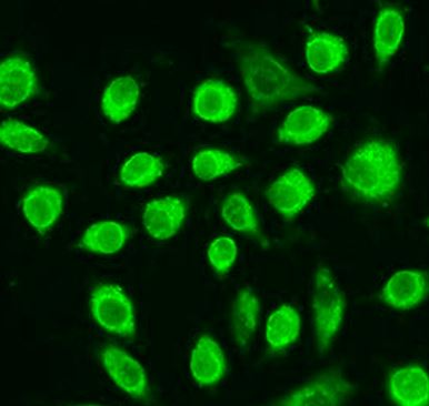
ARG43292 anti-GNAS antibody ICC/IF image
Immunofluorescence: L929 cells stained with ARG43292 anti-GNAS antibody at 1:100 dilution.
-
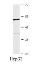
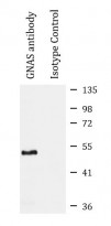
ARG43292 anti-GNAS antibody WB image
Western blot: 25 µg of HepG2 cell lysate stained with ARG43292 anti-GNAS antibody at 1:1000 dilution.
-
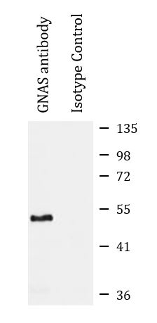
ARG43292 anti-GNAS antibody IP image
Immunoprecipitation: 200 µg extracts of Mouse brain cells were immunoprecipitated and stained with ARG43292 anti-GNAS antibody at 1:1000 dilution.