ARG11118
anti-GFAP antibody
anti-GFAP antibody for IHC-Frozen sections,Western blot and Human,Mouse,Rat,Cow
Overview
| Product Description | Goat Polyclonal antibody recognizes GFAP |
|---|---|
| Tested Reactivity | Hu, Ms, Rat, Cow |
| Tested Application | IHC-Fr, WB |
| Host | Goat |
| Clonality | Polyclonal |
| Isotype | IgG |
| Target Name | GFAP |
| Antigen Species | Human |
| Immunogen | Recombinant full-length Human GFAP isotype 1. |
| Conjugation | Un-conjugated |
| Alternate Names | Glial fibrillary acidic protein; ALXDRD; GFAP |
Application Instructions
| Application Suggestion |
|
||||||
|---|---|---|---|---|---|---|---|
| Application Note | * The dilutions indicate recommended starting dilutions and the optimal dilutions or concentrations should be determined by the scientist. |
Properties
| Form | Liquid |
|---|---|
| Purification | Affinity purified. |
| Buffer | PBS, 5 mM Sodium azide and 50% Glycerol. |
| Preservative | 5 mM Sodium azide |
| Stabilizer | 50% Glycerol |
| Concentration | 1 mg/ml |
| Storage Instruction | For continuous use, store undiluted antibody at 2-8°C for up to a week. For long-term storage, aliquot and store at -20°C. Storage in frost free freezers is not recommended. Avoid repeated freeze/thaw cycles. Suggest spin the vial prior to opening. The antibody solution should be gently mixed before use. |
| Note | For laboratory research only, not for drug, diagnostic or other use. |
Bioinformation
| Database Links | |
|---|---|
| Gene Symbol | GFAP |
| Gene Full Name | glial fibrillary acidic protein |
| Background | This gene encodes one of the major intermediate filament proteins of mature astrocytes. It is used as a marker to distinguish astrocytes from other glial cells during development. Mutations in this gene cause Alexander disease, a rare disorder of astrocytes in the central nervous system. Alternative splicing results in multiple transcript variants encoding distinct isoforms. [provided by RefSeq, Oct 2008] |
| Function | GFAP, a class-III intermediate filament, is a cell-specific marker that, during the development of the central nervous system, distinguishes astrocytes from other glial cells. [UniProt] |
| Cellular Localization | Cytoplasm. Note=Associated with intermediate filaments. [UniProt] |
| Calculated MW | 50 kDa |
| PTM | Phosphorylated by PKN1. [UniProt] |
Images (2) Click the Picture to Zoom In
-
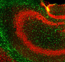
ARG11118 anti-GFAP antibody IHC-Fr image
Immunohistochemistry: Frozen section of Mouse hippocampus tissue stained with ARG11118 anti-GFAP antibody (green) at 1:5000 dilution, and co-stained with ARG52283 anti-FOX3 / NeuN antibody [1B7] (red) at 1:2000 dilution. Hoechst (blue) for nuclear staining. (Sample preparation: Following transcardial perfusion of mouse with 4% paraformaldehyde, brain was post fixed for 24 hours, cut to 45 µM, and free-floating sections were stained with above antibodies.).
-
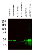
ARG11118 anti-GFAP antibody WB image
Western blot: Rat cortex, Rat cerebellum, Mouse cortex, Mouse cerebellum, Cow cortex and Cow cerebellum lysates stained with ARG11118 anti-GFAP antibody at 1:5000 dilution.
Strong band at about 50 kDa corresponds to GFAP protein. Smaller proteolytic fragments of GFAP are also detected on the blot.


