ARG63863
anti-GATA1 antibody
anti-GATA1 antibody for Flow cytometry,ICC/IF,Western blot and Human,Mouse
Developmental Biology antibody; Gene Regulation antibody
Overview
| Product Description | Goat Polyclonal antibody recognizes GATA1 |
|---|---|
| Tested Reactivity | Hu, Ms |
| Predict Reactivity | Cow, Rat, Dog |
| Tested Application | FACS, ICC/IF, WB |
| Host | Goat |
| Clonality | Polyclonal |
| Isotype | IgG |
| Target Name | GATA1 |
| Antigen Species | Human |
| Immunogen | C-DAEAYRHSPVFQ |
| Conjugation | Un-conjugated |
| Alternate Names | XLTDA; Eryf1; GATA-1; GF-1; GF1; NF-E1; ERYF1; XLANP; NFE1; GATA-binding factor 1; XLTT; NF-E1 DNA-binding protein; Erythroid transcription factor |
Application Instructions
| Application Suggestion |
|
||||||||
|---|---|---|---|---|---|---|---|---|---|
| Application Note | WB: Recommend incubate at RT for 1h. * The dilutions indicate recommended starting dilutions and the optimal dilutions or concentrations should be determined by the scientist. |
Properties
| Form | Liquid |
|---|---|
| Purification | Purified from goat serum by ammonium sulphate precipitation followed by antigen affinity chromatography using the immunizing peptide. |
| Buffer | Tris saline (pH 7.3), 0.02% Sodium azide and 0.5% BSA |
| Preservative | 0.02% Sodium azide |
| Stabilizer | 0.5% BSA |
| Concentration | 0.5 mg/ml |
| Storage Instruction | For continuous use, store undiluted antibody at 2-8°C for up to a week. For long-term storage, aliquot and store at -20°C or below. Storage in frost free freezers is not recommended. Avoid repeated freeze/thaw cycles. Suggest spin the vial prior to opening. The antibody solution should be gently mixed before use. |
| Note | For laboratory research only, not for drug, diagnostic or other use. |
Bioinformation
| Database Links | |
|---|---|
| Background | This gene encodes a protein which belongs to the GATA family of transcription factors. The protein plays an important role in erythroid development by regulating the switch of fetal hemoglobin to adult hemoglobin. Mutations in this gene have been associated with X-linked dyserythropoietic anemia and thrombocytopenia. [provided by RefSeq, Jul 2008] |
| Research Area | Developmental Biology antibody; Gene Regulation antibody |
| Calculated MW | 43 kDa |
| PTM | Highly phosphorylated on serine residues. Phosphorylation on Ser-310 is enhanced on erythroid differentiation. Phosphorylation on Ser-142 promotes sumoylation on Lys-137 (By similarity). Sumoylation on Lys-137 is enhanced by phosphorylation on Ser-142 and by interaction with PIAS4. Sumoylation with SUMO1 has no effect on transcriptional activity (By similarity). Acetylated at 2 conserved lysine-rich motifs by CREBBP in vitro. Acetylation does not affect DNA-binding in vitro but is essential to induce erythroid differentiation and for binding chromatin in vivo (By similarity). Acetylated on Lys-233, Lys-245 Lys-246 by EP300. |
Images (5) Click the Picture to Zoom In
-
ARG63863 anti-GATA1 antibody WB image
Western Blot: human PBMC lysate (35 µg protein in RIPA buffer) stained with ARG63863 anti-GATA1 antibody at 0.3 µg/ml dilution.
-

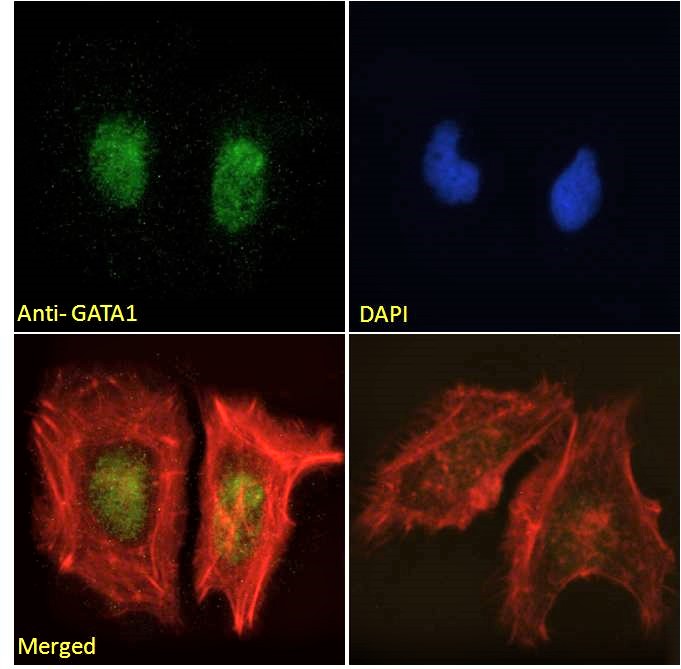
ARG63863 anti-GATA1 antibody ICC/IF image
Immunofluorescence: Paraformaldehyde fixed HeLa cells permeabilized with 0.15% Triton. Cells were stained with ARG63863 anti-GATA1 antibody (green) at 10 µg/ml dilution for 1 hour. DAPI (blue) for nuclear staining. Phalloidin (red) for Actin filaments staining. Negative control: Unimmunized goat IgG (green) at 10 µg/ml dilution.
-
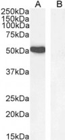
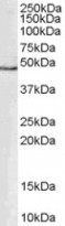
ARG63863 anti-GATA1 antibody WB image
Western blot: 35 µg of K562 nuclear lysate (A) and Human hippocampus (B, negative control) lysates (in RIPA buffer) stained with ARG63863 anti-GATA1 antibody at 1 µg/ml dilution and incubated at RT for 1 hour.
-

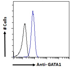
ARG63863 anti-GATA1 antibody FACS image
Flow Cytometry: Paraformaldehyde-fixed K562 cells permeabilized with 0.5% Triton. Cells were stained with ARG63863 anti-GATA1 antibody (blue line) at 10 µg/ml dilution for 1 hour, followed by incubation with Alexa FluorR 488 labelled secondary antibody. IgG control: Unimmunized goat IgG (black line).
-
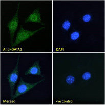
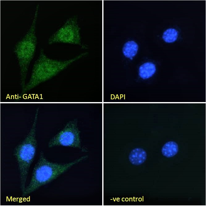
ARG63863 anti-GATA1 antibody ICC/IF image
Immunofluorescence: Paraformaldehyde fixed NIH/3T3 cells permeabilized with 0.15% Triton. Cells were stained with ARG63863 anti-GATA1 antibody (green) at 10 µg/ml dilution for 1 hour. DAPI (blue) for nuclear staining. Negative control: Unimmunized goat IgG (green) at 10 µg/ml dilution.