ARG63899
anti-GAPDH antibody
anti-GAPDH antibody for ICC/IF,IHC-Formalin-fixed paraffin-embedded sections,Western blot and Human,Mouse,Rat
Cancer antibody; Controls and Markers antibody; Metabolism antibody; Neuroscience antibody; Signaling Transduction antibody; Loading Control antibody; Loading Control antibody for Cytoplasmic Fractions; Organelle Marker antibody for Cytoplasm; Autophagy Study antibody
Overview
| Product Description | Goat Polyclonal antibody recognizes GAPDH |
|---|---|
| Tested Reactivity | Hu, Ms, Rat |
| Predict Reactivity | Dog |
| Tested Application | ICC/IF, IHC-P, WB |
| Specificity | GAPDH is constitutively expressed in almost all tissues at high levels. It is therefore a useful marker when a loading/positive control is required in western blotting. |
| Host | Goat |
| Clonality | Polyclonal |
| Isotype | IgG |
| Target Name | GAPDH |
| Antigen Species | Human |
| Immunogen | C-GVNHEKYDNSLK |
| Conjugation | Un-conjugated |
| Alternate Names | Glyceraldehyde-3-phosphate dehydrogenase; GAPD; HEL-S-162eP; G3PD; GAPDH; Peptidyl-cysteine S-nitrosylase GAPDH; EC 2.6.99.-; EC 1.2.1.12 |
Application Instructions
| Application Suggestion |
|
||||||||
|---|---|---|---|---|---|---|---|---|---|
| Application Note | IHC-P: Antigen Retrieval: Steam tissue section in Citrate buffer (pH 6.0). WB: Recommend incubate at RT for 1h. * The dilutions indicate recommended starting dilutions and the optimal dilutions or concentrations should be determined by the scientist. |
Properties
| Form | Liquid |
|---|---|
| Purification | Purified from goat serum by antigen affinity chromatography. |
| Buffer | Tris saline (pH 7.3), 0.02% Sodium azide and 0.5% BSA. |
| Preservative | 0.02% Sodium azide |
| Stabilizer | 0.5% BSA |
| Concentration | 0.5 mg/ml |
| Storage Instruction | For continuous use, store undiluted antibody at 2-8°C for up to a week. For long-term storage, aliquot and store at -20°C or below. Storage in frost free freezers is not recommended. Avoid repeated freeze/thaw cycles. Suggest spin the vial prior to opening. The antibody solution should be gently mixed before use. |
| Note | For laboratory research only, not for drug, diagnostic or other use. |
Bioinformation
| Database Links | |
|---|---|
| Gene Symbol | GAPDH |
| Gene Full Name | glyceraldehyde-3-phosphate dehydrogenase |
| Background | GAPDH protein has been identified as a moonlighting protein based on its ability to perform mechanistically distinct functions. The product of this gene catalyzes an important energy-yielding step in carbohydrate metabolism, the reversible oxidative phosphorylation of glyceraldehyde-3-phosphate in the presence of inorganic phosphate and nicotinamide adenine dinucleotide (NAD). The encoded protein has additionally been identified to have uracil DNA glycosylase activity in the nucleus. Also, this protein contains a peptide that has antimicrobial activity against E. coli, P. aeruginosa, and C. albicans. Studies of a similar protein in mouse have assigned a variety of additional functions including nitrosylation of nuclear proteins, the regulation of mRNA stability, and acting as a transferrin receptor on the cell surface of macrophage. Many pseudogenes similar to this locus are present in the human genome. Alternative splicing results in multiple transcript variants. [provided by RefSeq, Nov 2014] |
| Function | GAPDH has both glyceraldehyde-3-phosphate dehydrogenase and nitrosylase activities, thereby playing a role in glycolysis and nuclear functions, respectively. Participates in nuclear events including transcription, RNA transport, DNA replication and apoptosis. Nuclear functions are probably due to the nitrosylase activity that mediates cysteine S-nitrosylation of nuclear target proteins such as SIRT1, HDAC2 and PRKDC. Modulates the organization and assembly of the cytoskeleton. Facilitates the CHP1-dependent microtubule and membrane associations through its ability to stimulate the binding of CHP1 to microtubules. Glyceraldehyde-3-phosphate dehydrogenase is a key enzyme in glycolysis that catalyzes the first step of the pathway by converting D-glyceraldehyde 3-phosphate (G3P) into 3-phospho-D-glyceroyl phosphate. Component of the GAIT (gamma interferon-activated inhibitor of translation) complex which mediates interferon-gamma-induced transcript-selective translation inhibition in inflammation processes. Upon interferon-gamma treatment assembles into the GAIT complex which binds to stem loop-containing GAIT elements in the 3'-UTR of diverse inflammatory mRNAs (such as ceruplasmin) and suppresses their translation. [UniProt] |
| Research Area | Cancer antibody; Controls and Markers antibody; Metabolism antibody; Neuroscience antibody; Signaling Transduction antibody; Loading Control antibody; Loading Control antibody for Cytoplasmic Fractions; Organelle Marker antibody for Cytoplasm; Autophagy Study antibody |
| Calculated MW | 36 kDa |
| PTM | S-nitrosylation of Cys-152 leads to interaction with SIAH1, followed by translocation to the nucleus (By similarity). S-nitrosylation of Cys-247 is induced by interferon-gamma and LDL(ox) implicating the iNOS-S100A8/9 transnitrosylase complex and seems to prevent interaction with phosphorylated RPL13A and to interfere with GAIT complex activity. ISGylated. Sulfhydration at Cys-152 increases catalytic activity. Oxidative stress can promote the formation of high molecular weight disulfide-linked GAPDH aggregates, through a process called nucleocytoplasmic coagulation. Such aggregates can be observed in vivo in the affected tissues of patients with Alzheimer disease or alcoholic liver cirrhosis, or in cell cultures during necrosis. Oxidation at Met-46 may play a pivotal role in the formation of these insoluble structures. This modification has been detected in vitro following treatment with free radical donor (+/-)-(E)-4-ethyl-2-[(E)-hydroxyimino]-5-nitro-3-hexenamide. It has been proposed to destabilize nearby residues, increasing the likelihood of secondary oxidative damages, including oxidation of Tyr-45 and Met-105. This cascade of oxidations may augment GAPDH misfolding, leading to intermolecular disulfide cross-linking and aggregation. |
Images (6) Click the Picture to Zoom In
-
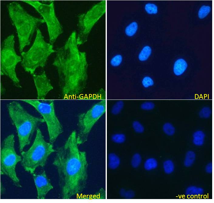
ARG63899 anti-GAPDH antibody ICC/IF image
Immunofluorescence: Paraformaldehyde fixed HeLa cells permeabilized with 0.15% Triton. Cells were stained with ARG63899 anti-GAPDH antibody (green) at 5 µg/ml dilution for 1 hour. DAPI (blue) for nuclear staining. Negative control: Unimmunized goat IgG (green) at 5 µg/ml dilution.
-

ARG63899 anti-GAPDH antibody IHC-P image
Immunohistochemistry: Paraffin-embedded Human liver tissue. Antigen Retrieval: Steam tissue section in Citrate buffer (pH 6.0). The tissue section was stained with ARG63899 anti-GAPDH antibody at 2 µg/ml dilution, followed by HRP-staining.
-

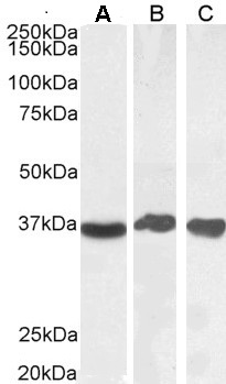
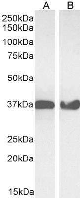
ARG63899 anti-GAPDH antibody WB image
Western blot: 35 µg of Human liver (A), Human testis (B) and Human tonsil (C) lysates (in RIPA buffer) stained with ARG63899 anti-GAPDH antibody at 0.1 µg/ml (A) and 0.03 µg/ml (B, C) dilutions and incubated at RT for 1 hour.
-

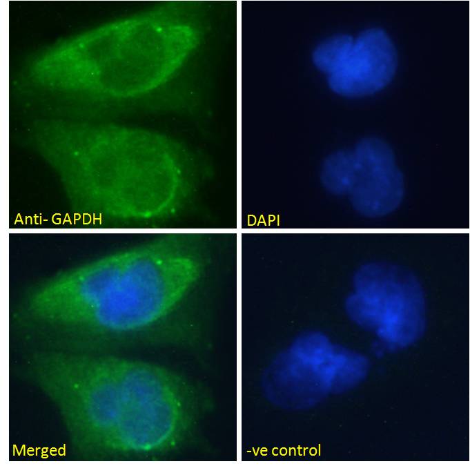
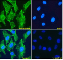
ARG63899 anti-GAPDH antibody ICC/IF image
Immunofluorescence: Paraformaldehyde fixed U251 cells permeabilized with 0.15% Triton. Cells were stained with ARG63899 anti-GAPDH antibody (green) at 10 µg/ml dilution for 1 hour. DAPI (blue) for nuclear staining. Negative control: Unimmunized goat IgG (green) at 10 µg/ml dilution.
-
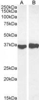
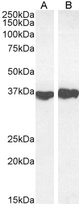
ARG63899 anti-GAPDH antibody WB image
Western blot: 35 µg of Mouse liver (A) and Rat heart (B) lysates (in RIPA buffer) stained with ARG63899 anti-GAPDH antibody at 0.1 µg/ml (A) and 0.03 µg/ml (B) dilutions and incubated at RT for 1 hour.
-

ARG63899 anti-GAPDH antibody WB image
Western blot: 35 µg of HeLa (A) and NIH/3T3 (B) cell lysates (in RIPA buffer) stained with ARG63899 anti-GAPDH antibody at 0.03 µg/ml dilution and incubated at RT for 1 hour.