ARG40828
anti-GALE antibody
anti-GALE antibody for IHC-Formalin-fixed paraffin-embedded sections,Western blot and Human,Mouse,Rat
Overview
| Product Description | Rabbit Polyclonal antibody recognizes GALE |
|---|---|
| Tested Reactivity | Hu, Ms, Rat |
| Tested Application | IHC-P, WB |
| Host | Rabbit |
| Clonality | Polyclonal |
| Isotype | IgG |
| Target Name | GALE |
| Antigen Species | Human |
| Immunogen | Recombinant protein corresponding to M1-N340 of Human GALE. |
| Conjugation | Un-conjugated |
| Alternate Names | UDP-GlcNAc 4-epimerase; SDR1E1; UDP-galactose 4-epimerase; Galactowaldenase; EC 5.1.3.2; EC 5.1.3.7; UDP-N-acetylglucosamine 4-epimerase; UDP-GalNAc 4-epimerase; UDP-N-acetylgalactosamine 4-epimerase; UDP-glucose 4-epimerase |
Application Instructions
| Application Suggestion |
|
||||||
|---|---|---|---|---|---|---|---|
| Application Note | IHC-P: Antigen Retrieval: Heat mediation was performed in Citrate buffer (pH 6.0, epitope retrieval solution) for 20 min. * The dilutions indicate recommended starting dilutions and the optimal dilutions or concentrations should be determined by the scientist. |
Properties
| Form | Liquid |
|---|---|
| Buffer | 0.2% Na2HPO4, 0.9% NaCl, 0.05% Sodium azide and 4% Trehalose. |
| Preservative | 0.05% Sodium azide |
| Stabilizer | 4% Trehalose |
| Concentration | 0.5 mg/ml |
| Storage Instruction | For continuous use, store undiluted antibody at 2-8°C for up to a week. For long-term storage, aliquot and store at -20°C or below. Storage in frost free freezers is not recommended. Avoid repeated freeze/thaw cycles. Suggest spin the vial prior to opening. The antibody solution should be gently mixed before use. |
| Note | For laboratory research only, not for drug, diagnostic or other use. |
Bioinformation
| Database Links | |
|---|---|
| Gene Symbol | GALE |
| Gene Full Name | UDP-galactose-4-epimerase |
| Background | This gene encodes UDP-galactose-4-epimerase which catalyzes two distinct but analogous reactions: the epimerization of UDP-glucose to UDP-galactose, and the epimerization of UDP-N-acetylglucosamine to UDP-N-acetylgalactosamine. The bifunctional nature of the enzyme has the important metabolic consequence that mutant cells (or individuals) are dependent not only on exogenous galactose, but also on exogenous N-acetylgalactosamine as a necessary precursor for the synthesis of glycoproteins and glycolipids. Mutations in this gene result in epimerase-deficiency galactosemia, also referred to as galactosemia type 3, a disease characterized by liver damage, early-onset cataracts, deafness and mental retardation, with symptoms ranging from mild ('peripheral' form) to severe ('generalized' form). Multiple alternatively spliced transcripts encoding the same protein have been identified. [provided by RefSeq, Jul 2008] |
| Function | Catalyzes two distinct but analogous reactions: the reversible epimerization of UDP-glucose to UDP-galactose and the reversible epimerization of UDP-N-acetylglucosamine to UDP-N-acetylgalactosamine. The reaction with UDP-Gal plays a critical role in the Leloir pathway of galactose catabolism in which galactose is converted to the glycolytic intermediate glucose 6-phosphate. It contributes to the catabolism of dietary galactose and enables the endogenous biosynthesis of both UDP-Gal and UDP-GalNAc when exogenous sources are limited. Both UDP-sugar interconversions are important in the synthesis of glycoproteins and glycolipids. [UniProt] |
| Calculated MW | 38 kDa |
Images (7) Click the Picture to Zoom In
-
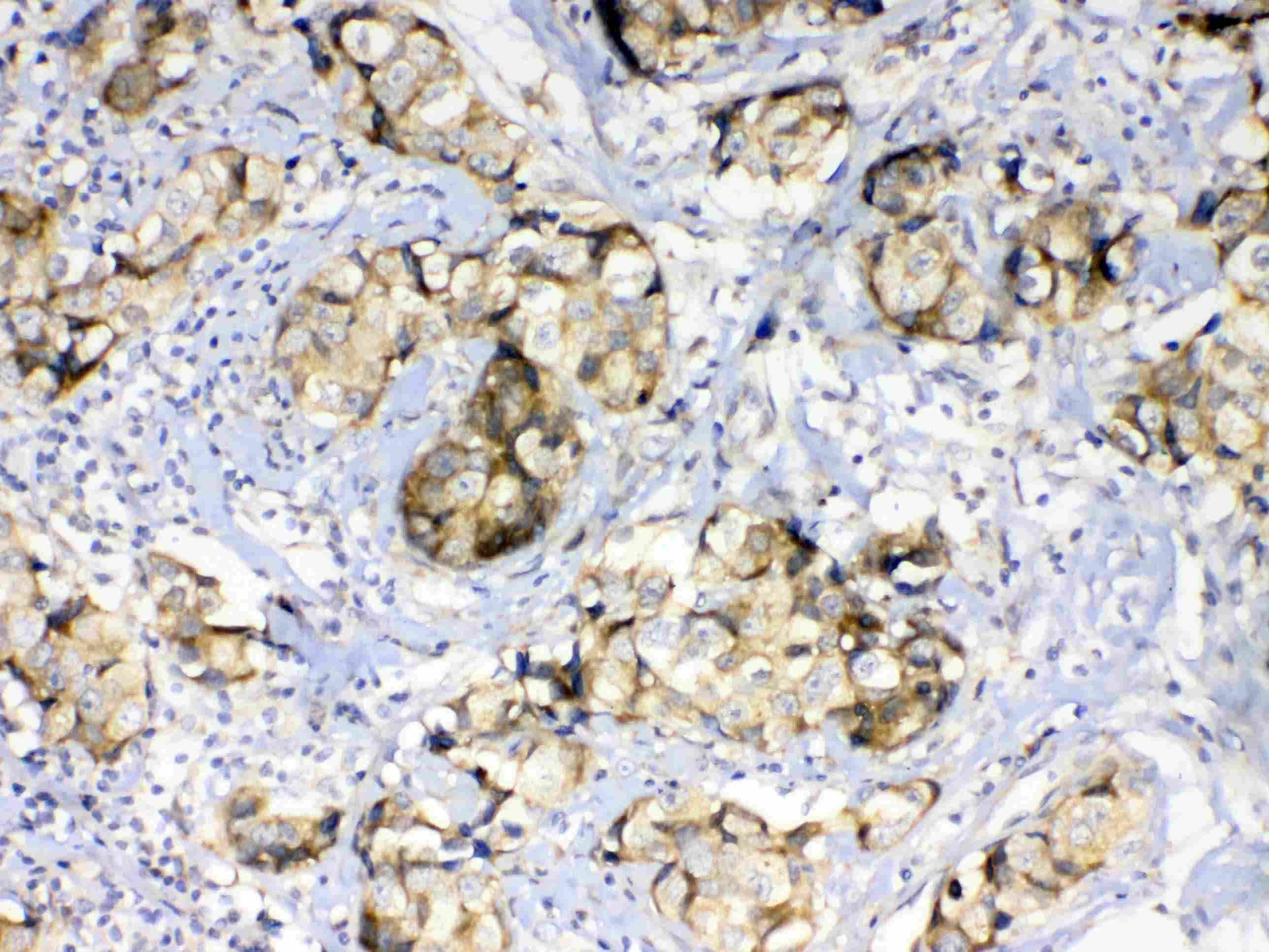
ARG40828 anti-GALE antibody IHC-P image
Immunohistochemistry: Paraffin-embedded Human mammary cancer tissue. Antigen Retrieval: Heat mediation was performed in Citrate buffer (pH 6.0, epitope retrieval solution) for 20 min. The tissue section was blocked with 10% goat serum. The tissue section was then stained with ARG40828 anti-GALE antibody at 1 µg/ml, overnight at 4°C.
-
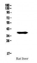
ARG40828 anti-GALE antibody WB image
Western blot: 50 µg of sample under reducing conditions. Rat liver lysate stained with ARG40828 anti-GALE antibody at 0.5 µg/ml, overnight at 4°C.
-

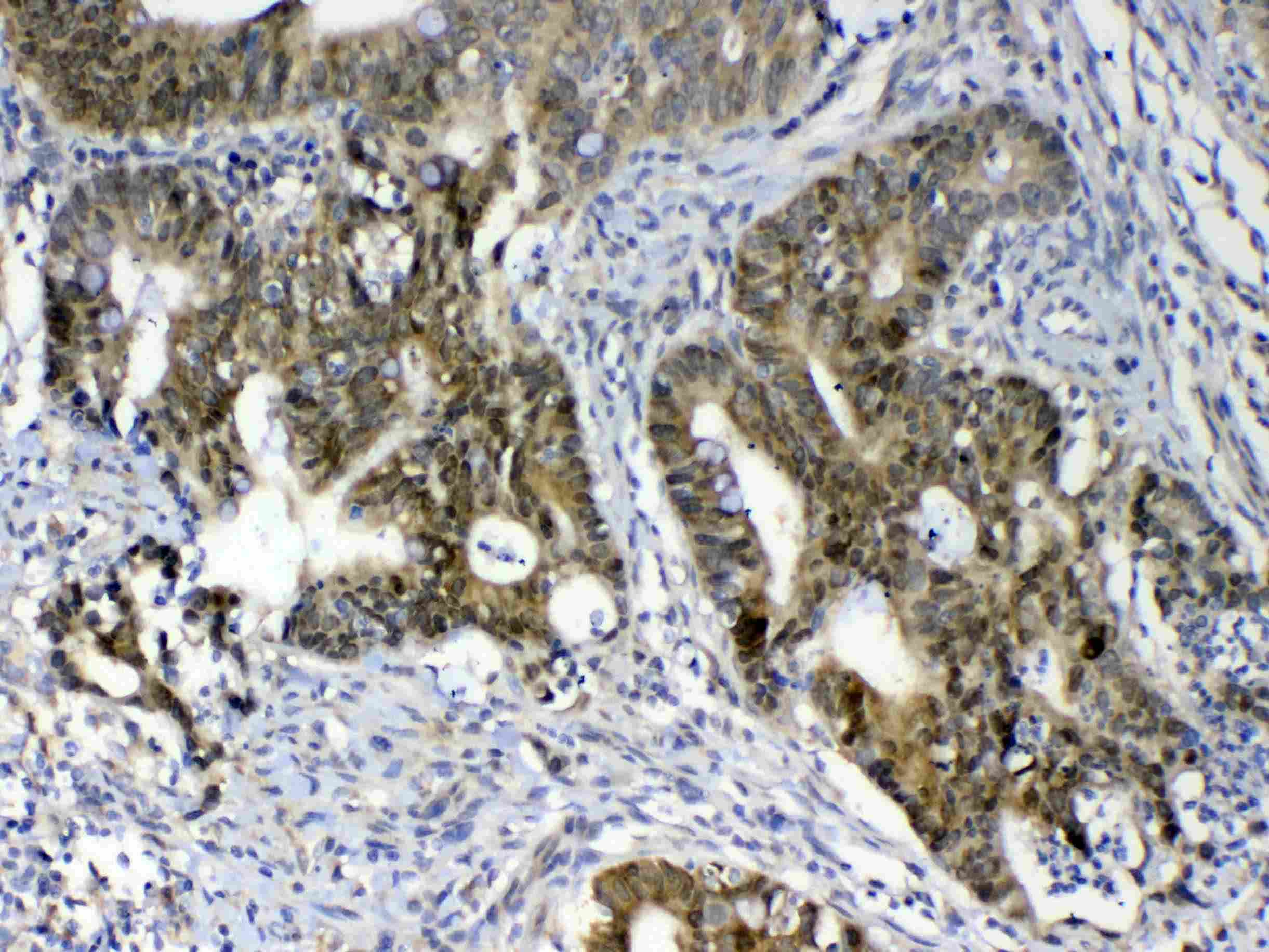
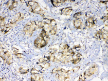
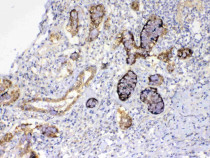
ARG40828 anti-GALE antibody IHC-P image
Immunohistochemistry: Paraffin-embedded Human colon cancer tissue. Antigen Retrieval: Heat mediation was performed in Citrate buffer (pH 6.0, epitope retrieval solution) for 20 min. The tissue section was blocked with 10% goat serum. The tissue section was then stained with ARG40828 anti-GALE antibody at 1 µg/ml, overnight at 4°C.
-
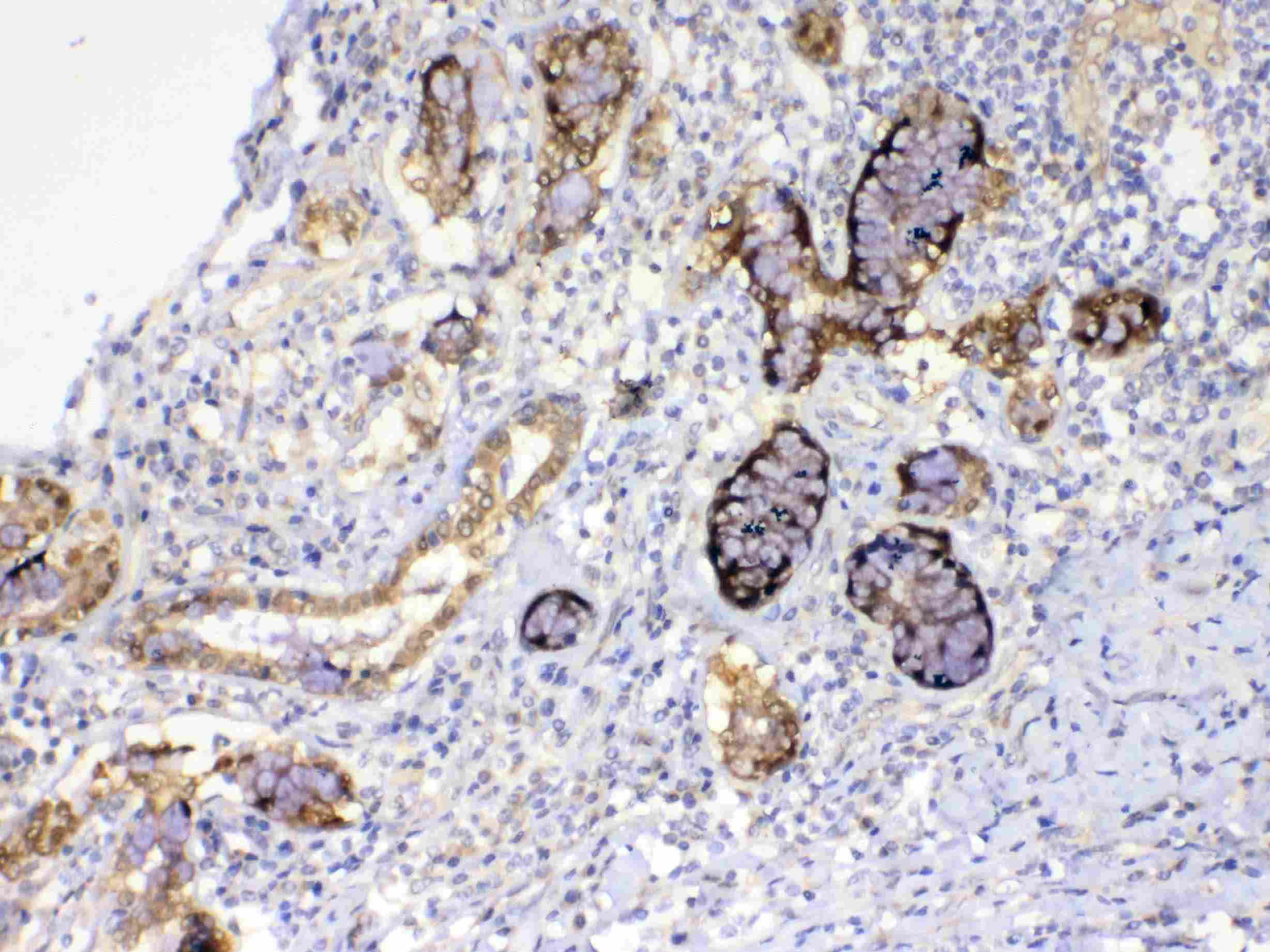
ARG40828 anti-GALE antibody IHC-P image
Immunohistochemistry: Paraffin-embedded Human lung cancer tissue. Antigen Retrieval: Heat mediation was performed in Citrate buffer (pH 6.0, epitope retrieval solution) for 20 min. The tissue section was blocked with 10% goat serum. The tissue section was then stained with ARG40828 anti-GALE antibody at 1 µg/ml, overnight at 4°C.
-
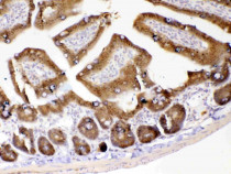
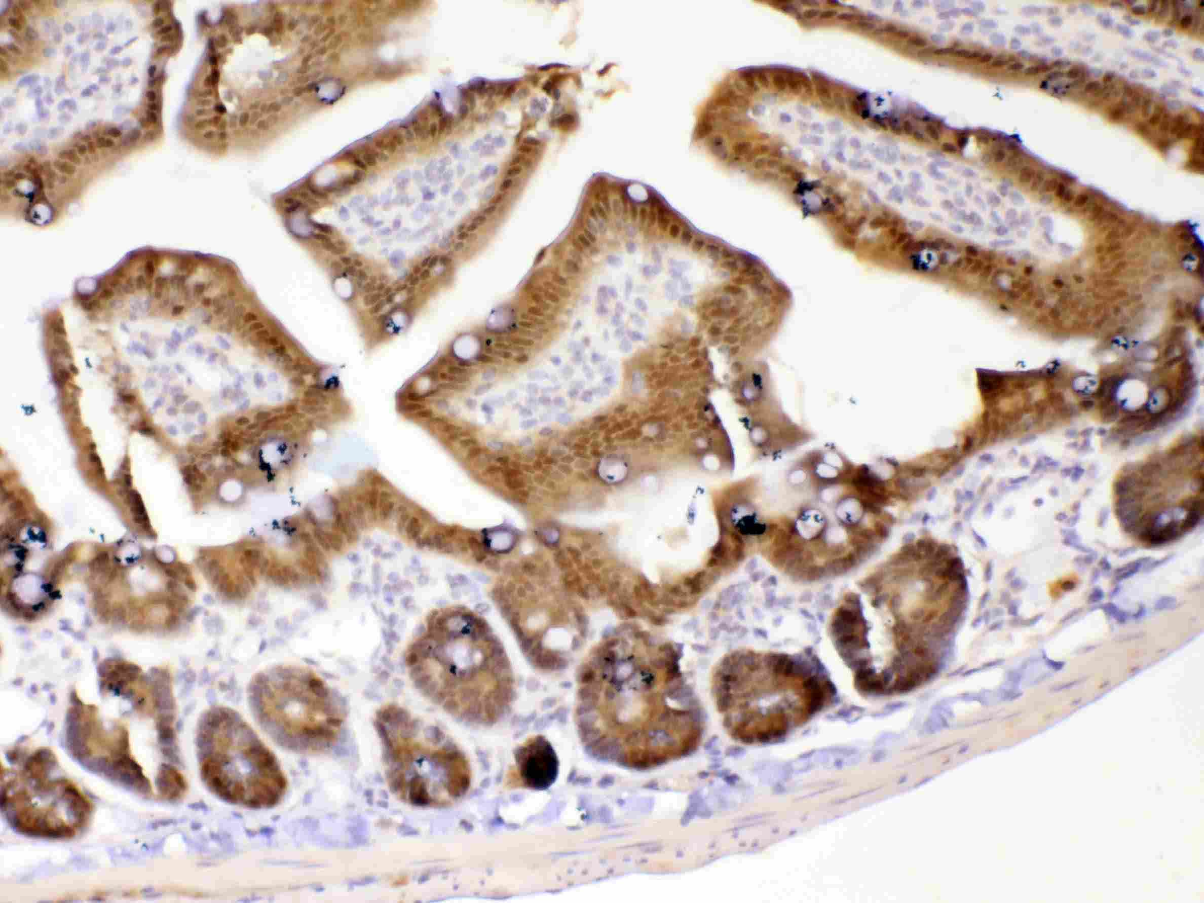
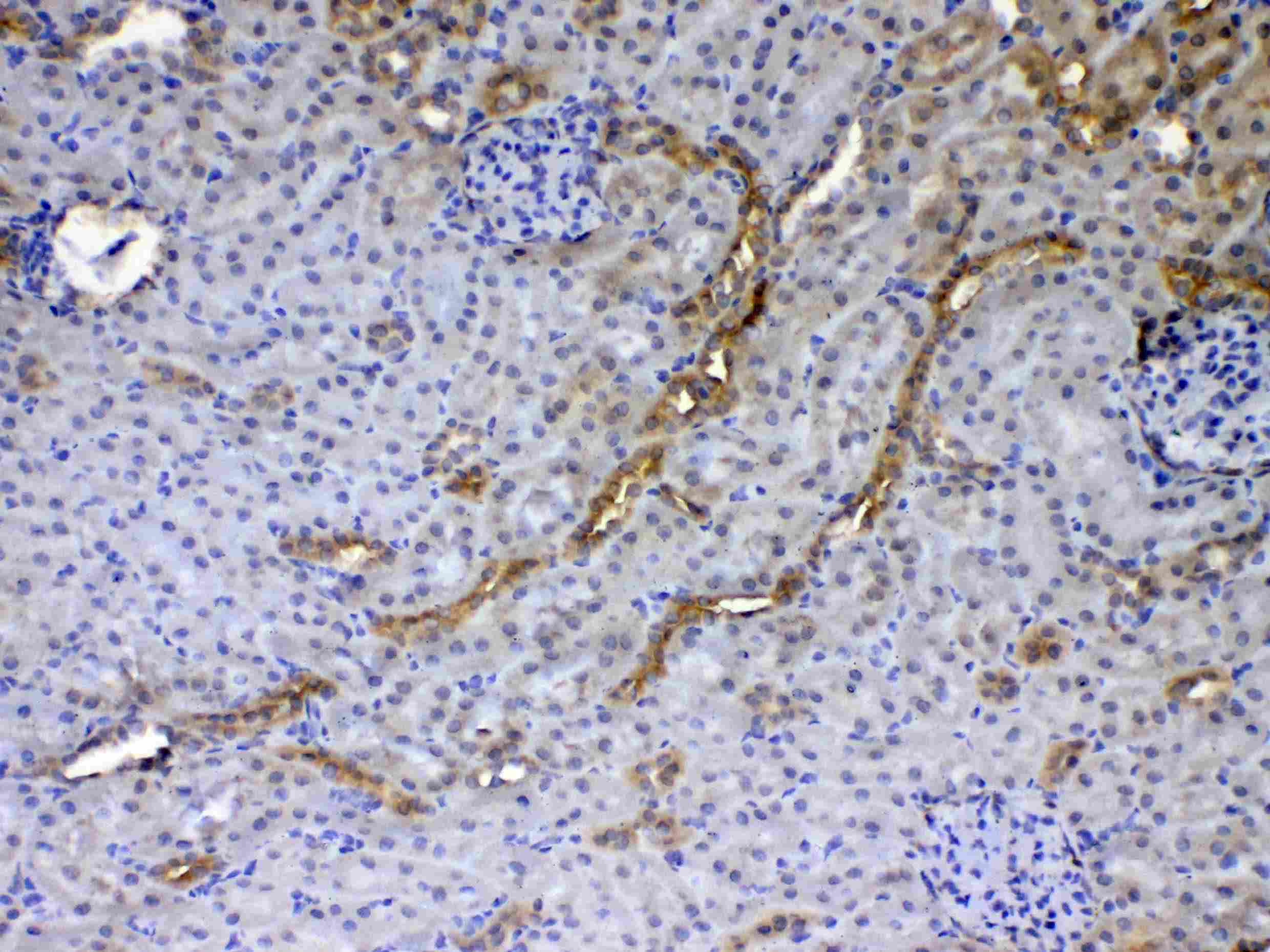
ARG40828 anti-GALE antibody IHC-P image
Immunohistochemistry: Paraffin-embedded Mouse intestine tissue. Antigen Retrieval: Heat mediation was performed in Citrate buffer (pH 6.0, epitope retrieval solution) for 20 min. The tissue section was blocked with 10% goat serum. The tissue section was then stained with ARG40828 anti-GALE antibody at 1 µg/ml, overnight at 4°C.
-

ARG40828 anti-GALE antibody IHC-P image
Immunohistochemistry: Paraffin-embedded Rat kidney tissue. Antigen Retrieval: Heat mediation was performed in Citrate buffer (pH 6.0, epitope retrieval solution) for 20 min. The tissue section was blocked with 10% goat serum. The tissue section was then stained with ARG40828 anti-GALE antibody at 1 µg/ml, overnight at 4°C.
-

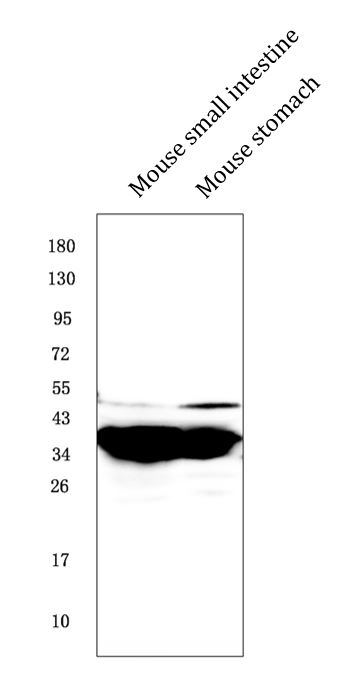
ARG40828 anti-GALE antibody WB image
Western blot: 50 µg of samples under reducing conditions. Mouse small intestine and Mouse stomach lysates stained with ARG40828 anti-GALE antibody at 0.5 µg/ml, overnight at 4°C.