ARG22243
anti-GABAB Receptor 1 antibody [S93A-49]
anti-GABAB Receptor 1 antibody [S93A-49] for ICC/IF,Western blot and Human,Mouse,Rat
Overview
| Product Description | Mouse Monoclonal antibody [S93A-49] recognizes GABAB Receptor 1 |
|---|---|
| Tested Reactivity | Hu, Ms, Rat |
| Tested Application | ICC/IF, WB |
| Specificity | Detects ~115kDa. No cross-reactivity against GABA(B)R2. |
| Host | Mouse |
| Clonality | Monoclonal |
| Clone | S93A-49 |
| Isotype | IgG1 |
| Target Name | GABAB Receptor 1 |
| Antigen Species | Rat |
| Immunogen | Fusion protein around aa. 873-977 (cytoplasmic C-terminus) of Rat GABA B Receptor 1 |
| Conjugation | Un-conjugated |
| Alternate Names | Gb1; GABA-B-R1; GABA-B receptor 1; GABA-BR1; GPRC3A; GABBR1-3; dJ271M21.1.2; dJ271M21.1.1; GABABR1; Gamma-aminobutyric acid type B receptor subunit 1; GB1 |
Application Instructions
| Application Suggestion |
|
||||||
|---|---|---|---|---|---|---|---|
| Application Note | * The dilutions indicate recommended starting dilutions and the optimal dilutions or concentrations should be determined by the scientist. |
Properties
| Form | Liquid |
|---|---|
| Purification | Purification with Protein G. |
| Buffer | PBS (pH 7.4), 0.09% Sodium azide and 50% Glycerol |
| Preservative | 0.09% Sodium azide |
| Stabilizer | 50% Glycerol |
| Concentration | 1 mg/ml |
| Storage Instruction | For continuous use, store undiluted antibody at 2-8°C for up to a week. For long-term storage, aliquot and store at -20°C. Storage in frost free freezers is not recommended. Avoid repeated freeze/thaw cycles. Suggest spin the vial prior to opening. The antibody solution should be gently mixed before use. |
| Note | For laboratory research only, not for drug, diagnostic or other use. |
Bioinformation
| Database Links | |
|---|---|
| Gene Symbol | Gabbr1 |
| Gene Full Name | gamma-aminobutyric acid (GABA) B receptor 1 |
| Background | Gamma-aminobutyric acid (GABA) is the main inhibitory neurotransmitter in the mammalian central nervous system. GABA exerts its effects through ionotropic [GABA(A/C)] receptors, to produce fast synaptic inhibition, and metabotropic [GABA(B)] receptors, to produce slow, prolonged inhibitory signals. The GABA(B) receptor consists of a heterodimer of two related 7-transmembrane receptors, GABA(B) receptor 1 and GABA(B) receptor 2. The GABA(B) receptor 1 gene is mapped to chromosome 6p21.3 within the HLA class I region close to the HLA-F gene. Susceptibility loci for multiple sclerosis, epilepsy, and schizophrenia have also been mapped in this region. Alternative splicing of this gene generates multiple transcript variants. [provided by RefSeq, Jun 2009] |
| Function | Component of a heterodimeric G-protein coupled receptor for GABA, formed by GABBR1 and GABBR2. Within the heterodimeric GABA receptor, only GABBR1 seems to bind agonists, while GABBR2 mediates coupling to G proteins. Ligand binding causes a conformation change that triggers signaling via guanine nucleotide-binding proteins (G proteins) and modulates the activity of down-stream effectors, such as adenylate cyclase. Signaling inhibits adenylate cyclase, stimulates phospholipase A2, activates potassium channels, inactivates voltage-dependent calcium-channels and modulates inositol phospholipid hydrolysis. Calcium is required for high affinity binding to GABA. Plays a critical role in the fine-tuning of inhibitory synaptic transmission. Pre-synaptic GABA receptor inhibits neurotransmitter release by down-regulating high-voltage activated calcium channels, whereas postsynaptic GABA receptor decreases neuronal excitability by activating a prominent inwardly rectifying potassium (Kir) conductance that underlies the late inhibitory postsynaptic potentials. Not only implicated in synaptic inhibition but also in hippocampal long-term potentiation, slow wave sleep, muscle relaxation and antinociception. Activated by (-)-baclofen, cgp27492 and blocked by phaclofen. Isoform 1E may regulate the formation of functional GABBR1/GABBR2 heterodimers by competing for GABBR2 binding. This could explain the observation that certain small molecule ligands exhibit differential affinity for central versus peripheral sites. [UniProt] |
| Cellular Localization | Cell Junction, Cell membrane, postsynaptic cell membrane, Synapse |
| Calculated MW | 108 kDa |
Images (2) Click the Picture to Zoom In
-
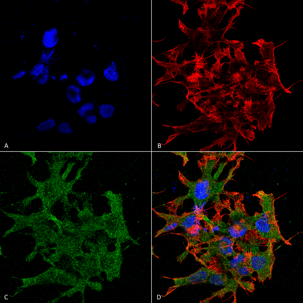
ARG22243 anti-GABAB Receptor 1 antibody [S93A-49] ICC/IF image
Immunofluorescence: Human Neuroblastoma cell line SK-N-BE. Fixation: 4% Formaldehyde for 15 min at RT. Primary antibody: ARG22243 anti-GABAB Receptor 1 antibody [S93A-49] at 1:100 for 60 min at RT. Secondary antibody: Goat anti-Mouse ATTO 488 at 1:100 for 60 min at RT. Counterstain: Phalloidin Texas Red F-Actin stain; DAPI (blue) nuclear stain. Magnification: 60X. (A) DAPI (blue) nuclear stain (B) Phalloidin Texas Red F-Actin stain (C) Primary antibody (D) Composite.
-
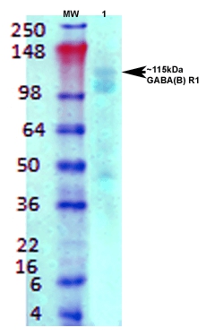
ARG22243 anti-GABAB Receptor 1 antibody [S93A-49] WB image
Western blot: Rat brain membrane lysate stained with ARG22243 anti-GABAB Receptor 1 antibody [S93A-49] at 1:1000 dilution.