ARG40876
anti-Furin antibody
anti-Furin antibody for ICC/IF,IHC-Formalin-fixed paraffin-embedded sections,Western blot and Human,Mouse,Rat
Overview
| Product Description | Rabbit Polyclonal antibody recognizes Furin |
|---|---|
| Tested Reactivity | Hu, Ms, Rat |
| Tested Application | ICC/IF, IHC-P, WB |
| Host | Rabbit |
| Clonality | Polyclonal |
| Isotype | IgG |
| Target Name | Furin |
| Antigen Species | Human |
| Immunogen | Synthetic peptide derived from Human Furin. |
| Conjugation | Un-conjugated |
| Alternate Names | FUR; Paired basic amino acid residue-cleaving enzyme; PCSK3; EC 3.4.21.75; PACE; Dibasic-processing enzyme; SPC1; Furin |
Application Instructions
| Application Suggestion |
|
||||||||
|---|---|---|---|---|---|---|---|---|---|
| Application Note | * The dilutions indicate recommended starting dilutions and the optimal dilutions or concentrations should be determined by the scientist. |
Properties
| Form | Liquid |
|---|---|
| Purification | Affinity purified. |
| Buffer | PBS (pH 7.4), 150 mM NaCl, 0.02% Sodium azide and 50% Glycerol. |
| Preservative | 0.02% Sodium azide |
| Stabilizer | 50% Glycerol |
| Storage Instruction | For continuous use, store undiluted antibody at 2-8°C for up to a week. For long-term storage, aliquot and store at -20°C. Storage in frost free freezers is not recommended. Avoid repeated freeze/thaw cycles. Suggest spin the vial prior to opening. The antibody solution should be gently mixed before use. |
| Note | For laboratory research only, not for drug, diagnostic or other use. |
Bioinformation
| Database Links | |
|---|---|
| Gene Symbol | FURIN |
| Gene Full Name | furin (paired basic amino acid cleaving enzyme) |
| Background | This gene encodes a member of the subtilisin-like proprotein convertase family, which includes proteases that process protein and peptide precursors trafficking through regulated or constitutive branches of the secretory pathway. It encodes a type 1 membrane bound protease that is expressed in many tissues, including neuroendocrine, liver, gut, and brain. The encoded protein undergoes an initial autocatalytic processing event in the ER and then sorts to the trans-Golgi network through endosomes where a second autocatalytic event takes place and the catalytic activity is acquired. The product of this gene is one of the seven basic amino acid-specific members which cleave their substrates at single or paired basic residues. Some of its substrates include proparathyroid hormone, transforming growth factor beta 1 precursor, proalbumin, pro-beta-secretase, membrane type-1 matrix metalloproteinase, beta subunit of pro-nerve growth factor and von Willebrand factor. It is also thought to be one of the proteases responsible for the activation of HIV envelope glycoproteins gp160 and gp140 and may play a role in tumor progression. This gene is located in close proximity to family member proprotein convertase subtilisin/kexin type 6 and upstream of the FES oncogene. Alternative splicing results in multiple transcript variants. [provided by RefSeq, Jan 2014] |
| Function | Furin is likely to represent the ubiquitous endoprotease activity within constitutive secretory pathways and capable of cleavage at the RX(K/R)R consensus motif. [UniProt] |
| Cellular Localization | Golgi apparatus, trans-Golgi network membrane; Single-pass type I membrane protein. Cell membrane; Single-pass type I membrane protein. Secreted. Endosome membrane; Single-pass type I membrane protein. Note=Shuttles between the trans-Golgi network and the cell surface. Propeptide cleavage is a prerequisite for exit of furin molecules out of the endoplasmic reticulum (ER). [UniProt] |
| Calculated MW | 87 kDa |
| PTM | The inhibition peptide, which plays the role of an intramolecular chaperone, is autocatalytically removed in the endoplasmic reticulum (ER) and remains non-covalently bound to furin as a potent autoinhibitor. Following transport to the trans Golgi, a second cleavage within the inhibition propeptide results in propeptide dissociation and furin activation. Phosphorylation is required for TGN localization of the endoprotease. In vivo, exists as di-, mono- and non-phosphorylated forms. [UniProt] |
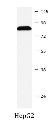
Images (1) Click the Picture to Zoom In
-

ARG40876 anti-Furin antibody WB image
Western blot: HepG2 cell lysate stained with ARG40876 anti-Furin antibody.