ARG10678
anti-Fibrillarin antibody
anti-Fibrillarin antibody for ICC/IF,IHC-Frozen sections,Western blot and Human,Mouse,Rat
Gene Regulation antibody; Nucleolar Marker antibody; DFC Marker antibody; Dense fibrillar component Marker antibody
Overview
| Product Description | Chicken Polyclonal antibody recognizes Fibrillarin |
|---|---|
| Tested Reactivity | Hu, Ms, Rat |
| Tested Application | ICC/IF, IHC-Fr, WB |
| Host | Chicken |
| Clonality | Polyclonal |
| Isotype | IgY |
| Target Name | Fibrillarin |
| Antigen Species | Human |
| Immunogen | Full length Human Fibrillarin expressed in and purified from E. coli. |
| Conjugation | Un-conjugated |
| Alternate Names | rRNA 2'-O-methyltransferase fibrillarin; RNU3IP1; 34 kDa nucleolar scleroderma antigen; FIB; FLRN; EC 2.1.1.-; Histone-glutamine methyltransferase |
Application Instructions
| Application Suggestion |
|
||||||||
|---|---|---|---|---|---|---|---|---|---|
| Application Note | * The dilutions indicate recommended starting dilutions and the optimal dilutions or concentrations should be determined by the scientist. |
Properties
| Form | Liquid |
|---|---|
| Buffer | PBS and 0.02% Sodium azide. |
| Preservative | 0.02% Sodium azide |
| Storage Instruction | For continuous use, store undiluted antibody at 2-8°C for up to a week. For long-term storage, aliquot and store at -20°C or below. Storage in frost free freezers is not recommended. Avoid repeated freeze/thaw cycles. Suggest spin the vial prior to opening. The antibody solution should be gently mixed before use. |
| Note | For laboratory research only, not for drug, diagnostic or other use. |
Bioinformation
| Database Links | |
|---|---|
| Gene Symbol | FBL |
| Gene Full Name | fibrillarin |
| Background | This gene product is a component of a nucleolar small nuclear ribonucleoprotein (snRNP) particle thought to participate in the first step in processing preribosomal RNA. It is associated with the U3, U8, and U13 small nuclear RNAs and is located in the dense fibrillar component (DFC) of the nucleolus. The encoded protein contains an N-terminal repetitive domain that is rich in glycine and arginine residues, like fibrillarins in other species. Its central region resembles an RNA-binding domain and contains an RNP consensus sequence. Antisera from approximately 8% of humans with the autoimmune disease scleroderma recognize fibrillarin. [provided by RefSeq, Jul 2008] |
| Function | S-adenosyl-L-methionine-dependent methyltransferase that has the ability to methylate both RNAs and proteins. Involved in pre-rRNA processing by catalyzing the site-specific 2'-hydroxyl methylation of ribose moieties in pre-ribosomal RNA. Site specificity is provided by a guide RNA that base pairs with the substrate. Methylation occurs at a characteristic distance from the sequence involved in base pairing with the guide RNA. Also acts as a protein methyltransferase by mediating methylation of 'Gln-105' of histone H2A (H2AQ104me), a modification that impairs binding of the FACT complex and is specifically present at 35S ribosomal DNA locus. [UniProt] |
| Research Area | Gene Regulation antibody; Nucleolar Marker antibody; DFC Marker antibody; Dense fibrillar component Marker antibody |
| Calculated MW | 34 kDa |
| PTM | By homology to other fibrillarins, some or all of the N-terminal domain arginines are modified to asymmetric dimethylarginine (DMA). |
Images (4) Click the Picture to Zoom In
-
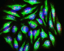
ARG10678 anti-Fibrillarin antibody ICC/IF image
Immunocytochemistry: HeLa cells stained with ARG10678 anti-Fibrillarin antibody which binds to nucleoli (red). Cells are also co-stained in green with monoclonal antibody to vimentin. DNA is revealed with DAPI.
-
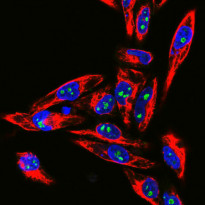
ARG10678 anti-Fibrillarin antibody ICC/IF image
Immunofluorescence: HeLa cells stained with ARG10678 anti-Fibrillarin antibody (green) at 1:10000 dilution and costained with ARG52469 anti-Vimentin antibody [2D1] (red) at 1:1000 dilution. DAPI (blue) for nuclear staining.
The Fibrillarin antibody stains nucleoli while the vimentin antibody binds to cytoplasmic intermediate filaments.
-
ARG10678 anti-Fibrillarin antibody WB image
Western blot: HeLa cell lysate stained with ARG10678 anti-Fibrillarin antibody.
-
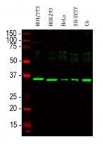
ARG10678 anti-Fibrillarin antibody WB image
Western blot: NIH/3T3, HEK293, HeLa, SH-SY5Y and C6 cell lysates stained with ARG10678 anti-Fibrillarin antibody at 1:5000 dilution.


