ARG63507
anti-FOXP3 antibody
anti-FOXP3 antibody for Flow cytometry,IHC-Formalin-fixed paraffin-embedded sections and Mouse
Cell Biology and Cellular Response antibody; Gene Regulation antibody; Immune System antibody; Regulatory T cells Study antibody
Overview
| Product Description | Goat Polyclonal antibody recognizes FOXP3 |
|---|---|
| Tested Reactivity | Ms |
| Predict Reactivity | Rat |
| Tested Application | FACS, IHC-P |
| Host | Goat |
| Clonality | Polyclonal |
| Isotype | IgG |
| Target Name | FOXP3 |
| Antigen Species | Mouse |
| Immunogen | KRSQRPNKCSNP |
| Conjugation | Un-conjugated |
| Alternate Names | FOXP3; Forkhead Box P3; DIETER; XPID; AIID; PIDX; IPEX; JM2; Immune Dysregulation, Polyendocrinopathy, Enteropathy, X-Linked; Forkhead Box Protein P3; SCURFIN; Immunodeficiency, Polyendocrinopathy, Enteropathy, X-Linked; FOXP3delta7; Scurfin |
Application Instructions
| Predict Reactivity Note | This antibody is predicted to react to rat FOXP3 based on the product citation paper. (https://doi.org/10.46235/1028-7221-1013-APO) | ||||||
|---|---|---|---|---|---|---|---|
| Application Suggestion |
|
||||||
| Application Note | IHC-P: Antigen Retrieval: Steam tissue section in Tris/EDTA buffer (pH 9.0). * The dilutions indicate recommended starting dilutions and the optimal dilutions or concentrations should be determined by the scientist. |
Properties
| Form | Liquid |
|---|---|
| Purification | Purified from goat serum by antigen affinity chromatography. |
| Buffer | Tris saline (pH 7.3), 0.02% Sodium azide and 0.5% BSA. |
| Preservative | 0.02% Sodium azide |
| Stabilizer | 0.5% BSA |
| Concentration | 0.5 mg/ml |
| Storage Instruction | For continuous use, store undiluted antibody at 2-8°C for up to a week. For long-term storage, aliquot and store at -20°C or below. Storage in frost free freezers is not recommended. Avoid repeated freeze/thaw cycles. Suggest spin the vial prior to opening. The antibody solution should be gently mixed before use. |
| Note | For laboratory research only, not for drug, diagnostic or other use. |
Bioinformation
| Database Links | |
|---|---|
| Gene Symbol | FOXP3 |
| Gene Full Name | forkhead box P3 |
| Background | The protein encoded by this gene is a member of the forkhead/winged-helix family of transcriptional regulators. Defects in this gene are the cause of immunodeficiency polyendocrinopathy, enteropathy, X-linked syndrome (IPEX), also known as X-linked autoimmunity-immunodeficiency syndrome. Alternatively spliced transcript variants encoding different isoforms have been identified. [provided by RefSeq, Jul 2008] |
| Function | Transcriptional regulator which is crucial for the development and inhibitory function of regulatory T-cells (Treg). [UniProt] Plays an essential role in maintaining homeostasis of the immune system by allowing the acquisition of full suppressive function and stability of the Treg lineage, and by directly modulating the expansion and function of conventional T-cells. [UniProt] |
| Cellular Localization | Cytoplasm, Nucleus. [UniProt] |
| Highlight | Related products: FOXP3 antibodies; FOXP3 Duos / Panels; Anti-Rabbit IgG secondary antibodies; Related news: Tumor-Infiltrating Lymphocytes (TILs) |
| Research Area | Cell Biology and Cellular Response antibody; Gene Regulation antibody; Immune System antibody; Regulatory T cells Study antibody |
| Calculated MW | 47 kDa |
| PTM | Acetylation, Isopeptide bond, Phosphoprotein, Ubl conjugation. [UniProt] |
Images (3) Click the Picture to Zoom In
-
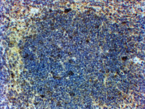
ARG63507 anti-FOXP3 antibody IHC-P image
Immunohistochemistry: Paraffin-embedded Mouse spleen tissue stained with ARG63507 anti-FOXP3 antibody at 2 - 4 µg/ml dilution.
-
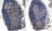
ARG63507 anti-FOXP3 antibody IHC-P image
Immunohistochemistry: Paraffin embedded Mouse Thymus (Right panel shows staining without primary antibody as negative control). (Steamed antigen retrieval with Tris/EDTA buffer pH 9) stained with ARG63507 anti-FOXP3 antibody at 2 µg/ml dilution followed by HRP-staining.
-
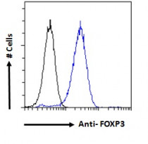
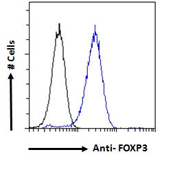
ARG63507 anti-FOXP3 antibody FACS image
Flow Cytometry: Paraformaldehyde-fixed NIH/3T3 cells permeabilized with 0.5% Triton. Cells were stained with ARG63507 anti-FOXP3 antibody (blue line) at 10 µg/ml dilution for 1 hour, followed by incubation with Alexa FluorR 488 labelled secondary antibody. IgG control: Unimmunized goat IgG (black line).


Specific References
Autoimmune profile of rat blood in experimental ulcerative colitis
IHC-P / Rat


