ARG10712
anti-FOX3 / NeuN antibody
anti-FOX3 / NeuN antibody for ICC/IF,IHC-Frozen sections,Western blot and Human,Mouse,Rat,Cow,Horse,Pig

Overview
| Product Description | Rabbit Polyclonal antibody recognizes FOX3 / NeuN |
|---|---|
| Tested Reactivity | Hu, Ms, Rat, Cow, Hrs, Pig |
| Predict Reactivity | Chk |
| Tested Application | ICC/IF, IHC-Fr, WB |
| Host | Rabbit |
| Clonality | Polyclonal |
| Isotype | IgG |
| Target Name | FOX3 / NeuN |
| Antigen Species | Human |
| Immunogen | N-terminal 100 aa. of Human Fox3 expressed in and purified from E. coli. |
| Conjugation | Un-conjugated |
| Alternate Names | RNA binding protein fox-1 homolog 3; NEUN; FOX-3; HRNBP3; Fox-1 homolog C; FOX3 |
Application Instructions
| Application Suggestion |
|
||||||||
|---|---|---|---|---|---|---|---|---|---|
| Application Note | * The dilutions indicate recommended starting dilutions and the optimal dilutions or concentrations should be determined by the scientist. |
Properties
| Form | Liquid |
|---|---|
| Purification | Affinity purification. |
| Buffer | PBS and 50% Glycerol. |
| Stabilizer | 50% Glycerol |
| Concentration | 1 mg/ml |
| Storage Instruction | For continuous use, store undiluted antibody at 2-8°C for up to a week. For long-term storage, aliquot and store at -20°C. Storage in frost free freezers is not recommended. Avoid repeated freeze/thaw cycles. Suggest spin the vial prior to opening. The antibody solution should be gently mixed before use. |
| Note | For laboratory research only, not for drug, diagnostic or other use. |
Bioinformation
| Database Links |
Swiss-port # A6NFN3 Human RNA binding protein fox-1 homolog 3 Swiss-port # Q8BIF2 Mouse RNA binding protein fox-1 homolog 3 |
|---|---|
| Gene Symbol | RBFOX3 |
| Gene Full Name | RNA binding protein, fox-1 homolog (C. elegans) 3 |
| Function | RNA-binding protein that regulates alternative splicing events. [UniProt] |
| Calculated MW | 34 kDa |
Images (4) Click the Picture to Zoom In
-
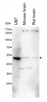
ARG10712 anti-FOX3 / NeuN antibody WB image
Western blot: 30 µg of U87, Mouse brain and Rat brain lysates stained with ARG10712 anti-FOX3 / NeuN antibody at 1:5000 dilution.
-
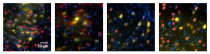
ARG10712 anti-FOX3 / NeuN antibody IHC-Fr image
Immunohistochemistry: Frozen section of Mouse C57BL/6Jnarl brain tissue. The tissue section was fixed by 4% formalin and blocked with BSA with 3% Goat serum, at RT for 1 hour. Tissue section was then stained with ARG10712 anti-FOX3 / NeuN antibody at 1:500 dilution, in PBS with 1% Goat serum, overnight at 4°C.
Blue: DAPI
Yellow: Venus reporter gene
Red: NueN -
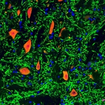
ARG10712 anti-FOX3 / NeuN antibody IHC-Fr image
Immunohistochemistry: Paraformaldehyde-fixed frozen section of adult Rat brain stem stained with ARG10712 anti-FOX3 / NeuN antibody (red) and co-stained for MAP2 (green). DNA is shown in blue. Note that RPCA-Fox3 stains neuronal nuclei and distal perikarya and that the MAP2 antibody stains the dendrites extending from these cells.
-
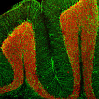
ARG10712 anti-FOX3 / NeuN antibody IHC-Fr image
Immunohistochemistry: Frozen section of Adult Mouse cerebellum stained with ARG10712 anti-FOX3 / NeuN antibody (red) at 1:5000 dilution and costained with ARG52313 anti-GFAP antibody (green) at 1:5000 dilution. (Sample preparation: Following transcardial perfusion with 4% paraformaldehyde, brain was post fixed for 24 hours and 45 µm free-floating sections were stained with above antibodies.)
The FOX3 / NeuN antibody stains the nuclei of neurons in the cerebellar granule layer. The GFAP antibody stains the processes of Bergmann glia in the molecular layer and astroglia in the granule and white matter layers.


Customer's Feedback
Good

Review for anti-FOX3 / NeuN antibody
Application:IHC
Sample:Mouse C57BL/6Jnarl brain tissue.
Sample Preparation Method:Frozen
Fixation Buffer:4% formalin
Primary Antibody Dilution Factor:1:500
Primary Antibody Incubation Time:overnight
Primary Antibody Incubation Temperature:4 ºC
Conjugation of Secondary Antibody:Dylight 594

Good

Review for anti-FOX3 / NeuN antibody
Application:WB
Sample:U87, Mouse brain and Rat brain
Sample Loading Amount:30 µg
Primary Antibody Dilution Factor:1:5000
Primary Antibody Incubation Time:overnight
Primary Antibody Incubation Temperature:4 ºC
Specific References
Comparison of Different Tissue Clearing Methods for Three-Dimensional Reconstruction of Human Brain Cellular Anatomy Using Advanced Imaging Techniques
IHC-Fr / Human


